. 2013 Jul 15;188(2):179-86.
doi: 10.1164/rccm.201211-2139OC.

Serum amyloid A promotes lung neutrophilia by increasing IL-17A levels in the mucosa and γδ T cells

Affiliations

Serum amyloid A promotes lung neutrophilia by increasing IL-17A levels in the mucosa and γδ T cells

Desiree Anthony et al. Am J Respir Crit Care Med. .

Abstract

Rationale: Neutrophilic inflammation is an important pathologic feature of chronic obstructive pulmonary disease (COPD) and infectious exacerbations of COPD. Serum amyloid A (SAA) promotes neutrophilic inflammation by its interaction with lung mucosal ALX/FPR2 receptors. However, little is known about how this endogenous mediator regulates IL-17A immunity.

Objectives: To determine whether SAA causes neutrophilic inflammation by IL-17A-dependent mechanisms.

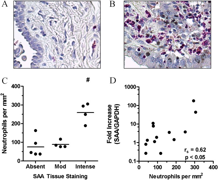
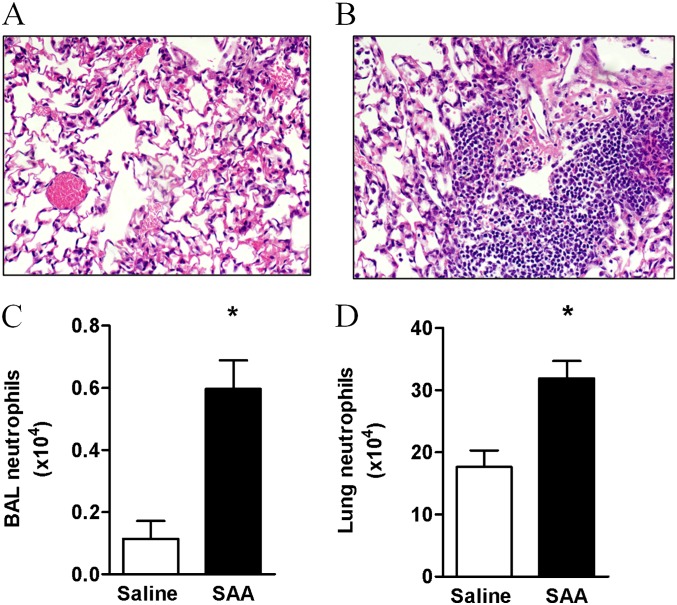
Methods: The relationship between SAA and neutrophils was investigated in lung sections from patients with COPD and a chronic mouse model of SAA exposure. A neutralizing antibody to IL-17A was used to block SAA responses in vivo, and a cell-sorting strategy was used to identify cellular sources.

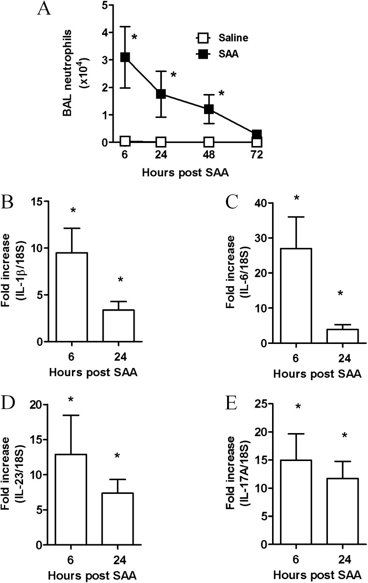
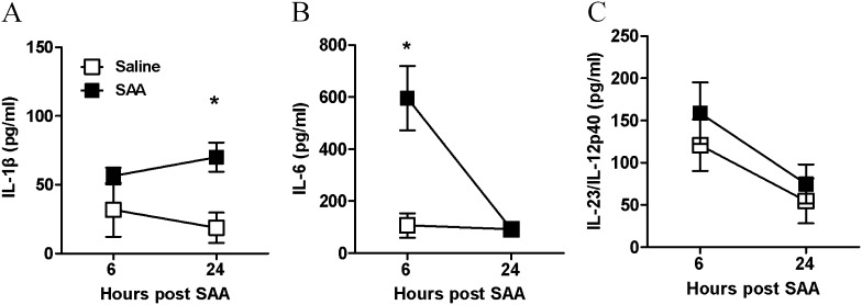
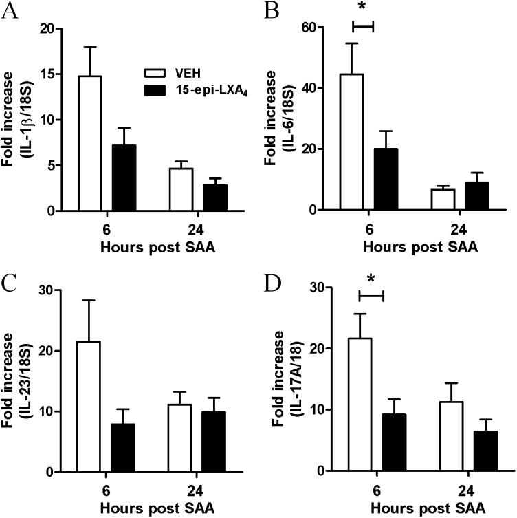
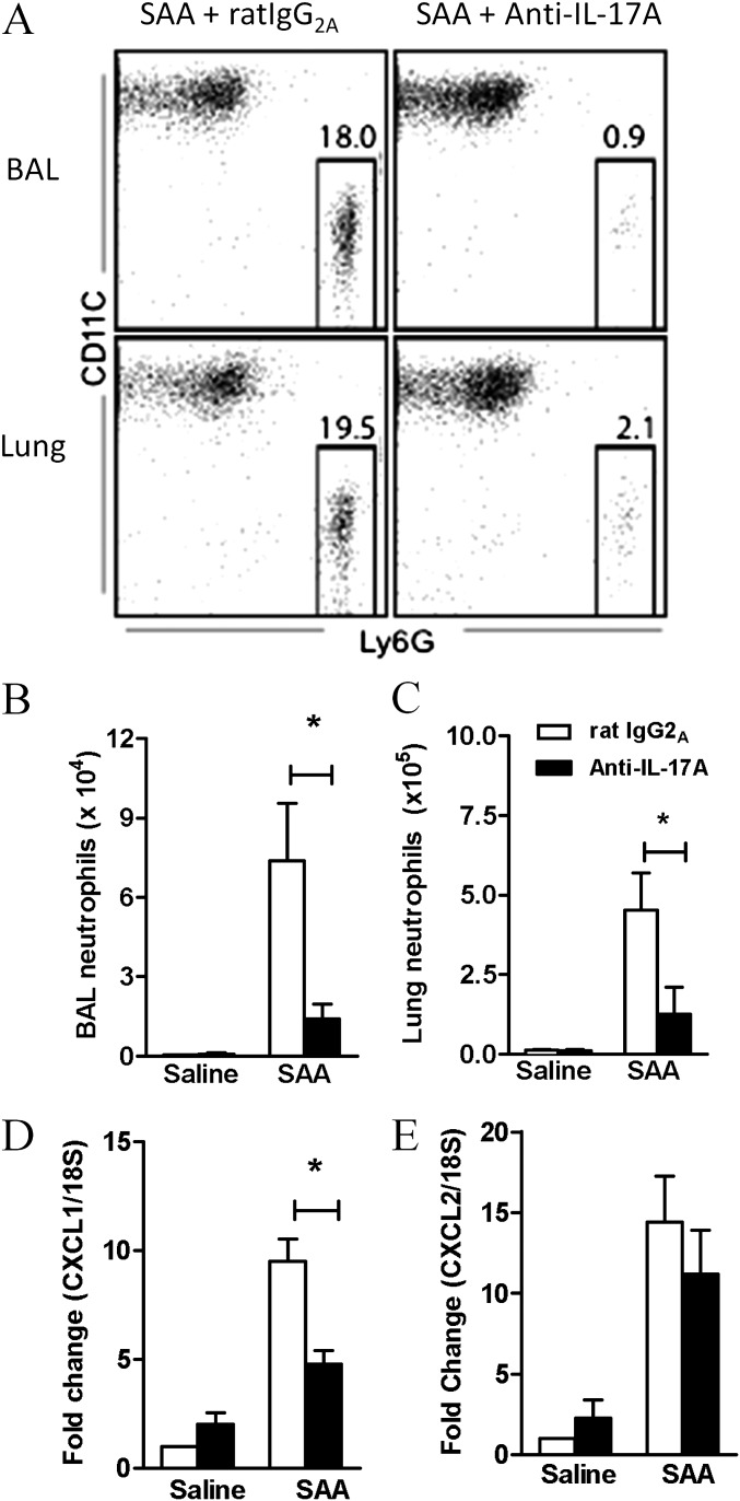
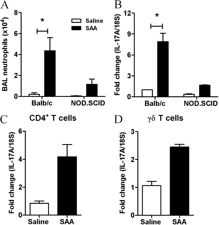
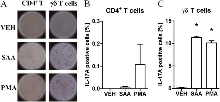
Measurements and main results: SAA mRNA expression was positively associated with tissue neutrophils in COPD (P < 0.05). SAA predominately promoted expression of the TH17 polarizing cytokine IL-6, which was opposed by 15-epi-lipoxin A4, a counter-regulatory mediator, and ALX/FPR2 ligand. SAA-induced inflammation was markedly reduced by a neutralizing antibody to IL-17A in vivo. Cellular sources of IL-17A induced by SAA include CD4(+) T cells, γδ T cells, and an Epcam(+)CD45(-) population enriched for epithelial cells. SAA promotes expression of IL-17A in γδ T cells and this innate cell proportionally expressed higher levels of IL-17A transcript than CD4(+) T cells or epithelial cells.

Conclusions: The SAA-IL-17A axis represents an important innate defense network that may underlie persistent neutrophilic airway inflammation in COPD and modulating the ALX/FPR2 receptor represents a novel approach to targeting aberrant IL-17A-mediated lung immunity.


Figures

<i>Figure 1.</i>
Figure 1.
The relationship between tissue neutrophils and serum amyloid A (SAA) levels in chronic obstructive pulmonary disease (COPD) lung. Representative tissue staining for the granulocyte marker esterase in (A) control and (B) COPD lung sections. (C) Esterase-positive tissue neutrophils were quantified and grouped according to the degree of SAA tissue staining, demonstrating a significant increase in neutrophils numbers in lung sections that displayed intense SAA tissue staining. (D) Correlation between SAA expression as determined by quantitative polymerase chain reaction and the number of neutrophils in serial COPD lung sections. n = 13 individual patient samples, #P < 0.05, Spearman correlation. GADPH = glyceraldehyde phosphate dehydrogenase.
<i>Figure 2.</i>
Figure 2.
Chronic administration of serum amyloid A (SAA) promotes neutrophil accumulation in the mouse lung. Mice were challenged with SAA (2 μg) or saline once a week for 5 weeks and lung tissue and bronchoalveolar lavage (BAL) fluid were harvested 24 hours after the final intranasal treatment of SAA. Representative hematoxylin and eosin staining of sections from (A) saline- or (B) SAA-treated mice. The hematoxylin and eosin staining demonstrates increased immune cell infiltrate in the lung sections after SAA treatment. The number of neutrophils was determined by fluorescence-activated cell sorter and pregated according to a single cell; viable (PI exclusion), large-granular cell gate and neutrophils were determined to be CD11C and Ly6G+ (Gr-1) in the (C) BAL and (D) lung. n = 3–4 mice, *P < 0.001.
<i>Figure 3.</i>
Figure 3.
Serum amyloid A (SAA)–mediated bronchoalveolar lavage (BAL) neutrophil recruitment is associated with expression of IL-17–related cytokine members in the lung. Mice were given a single dose of saline or SAA (2 μg) intranasally. BAL was performed and lungs were harvested at the indicated time points. (A) Total neutrophil numbers in the BAL fluid ± SEM, *P < 0.05. The mean ± SEM gene expression from three independent experiments (n = 5–8 mice per experimental time point) are presented. (B–E) Gene expression of IL-17A regulating cytokines, IL-6, IL-1β, IL-23, and IL-17A as determined by quantitative polymerase chain reaction, normalized to 18S and relative to naive mice. *P < 0.05.
<i>Figure 4.</i>
Figure 4.
Serum amyloid A (SAA) predominantly promotes protein secretion of the IL-17A–regulating cytokine, IL-6. Mice were treated intranasally with saline or SAA (2 μg) and bronchoalveolar lavage fluid collected at the indicated time points (hours). Bronchoalveolar lavage fluid concentrations of IL-17A–inducing cytokines (A) IL-1β, (B) IL-6, and (C) IL-23 after SAA treatment. n = 3–8 mice per group, *P < 0.05.
<i>Figure 5.</i>
Figure 5.
Serum amyloid A (SAA)–mediated expression of IL-17A and its related cytokines is dependent on ALX-FPR2. Mice were simultaneously challenged with SAA (2 μg) and 15-epi-LXA4 (4 μg) or vehicle by intranasal administration and gene expression of (A) IL-1β, (B) IL-6, (C) IL-23, and (D) IL-17A was determined in lung tissue at 6 or 24 hours. Quantitative polymerase chain reaction was used to determine gene expression relative to 18S. n = 4–5 mice per group, *P < 0.001.
<i>Figure 6.</i>
Figure 6.
Blocking IL-17A inhibits serum amyloid A (SAA)–induced neutrophil recruitment. Mice were pretreated with anti–IL-17A or rat IgG2A (isotype) antibody (50 μg) intranasally 1 hour before saline or SAA (2 μg) administration. (A) Representative fluorescence-activated cell sorter plot of each treatment group 24 hours after SAA administration. (B) Total number of neutrophils in the bronchoalveolar lavage (BAL) and (C) lung of treated mice. Pooled data from two independent experiments. n = 5–8 mice per group. Gene expression of the neutrophilic mediators (D) CXCL1 and (E) CXCL2 in the lung 6 hours after SAA administration. n = 4–5 mice, *P < 0.05.
<i>Figure 7.</i>
Figure 7.
Serum amyloid A (SAA)–induced IL-17A from multiple cellular sources. (A) Total bronchoalveolar lavage (BAL) neutrophils as determined by fluorescence-activated cell sorter in Balb/c and NOD.SCID mice treated with saline or SAA. (B) IL-17A gene expression in NOD.SCID or control mice 24 hours after intranasal SAA (2 μg) administration was determined by quantitative polymerase chain reaction. *P < 0.05. IL-17A gene expression from (C) CD4+T and (D) γδ T cells.
<i>Figure 8.</i>
Figure 8.
Enzyme-linked immunospot assay of the cellular source of IL-17A in response to serum amyloid A (SAA). Mice were intranasally treated with SAA and γδT (CD45+, γδTCR+), and CD4+T (TCR+, CD4+ TCR) cells were sorted. All cell populations were pregated according to single, viable (PI exclusion) cells. A total of 100,000 CD4+T or 500 γδT cells were stimulated with media alone (Veh), saline, SAA (1 μg/ml), or phorbol myristate acetate–ionomycin (PMA) for 24 hours. (A) Representative well of each treatment group shown. Percentage of IL-17A–producing (B) CD4+T and (C) γδT as determined by the number of positive spots divided by the total number of cells per well. Each sample was pooled from 8–10 mice. n = 2–3 wells per treatment, *P < 0.05.

References

    1. Hogg JC, Chu F, Utokaparch S, Woods R, Elliott WM, Buzatu L, Cherniack RM, Rogers RM, Sciurba FC, Coxson HO, et al. The nature of small-airway obstruction in chronic obstructive pulmonary disease. N Engl J Med. 2004;350:2645–2653. - PubMed
    1. Culpitt SV, Maziak W, Loukidis S, Nightingale JA, Matthews JL, Barnes PJ. Effect of high dose inhaled steroid on cells, cytokines, and proteases in induced sputum in chronic obstructive pulmonary disease. Am J Respir Crit Care Med. 1999;160:1635–1639. - PubMed
    1. Vlahos R, Wark PA, Anderson GP, Bozinovski S. Glucocorticosteroids differentially regulate MMP-9 and neutrophil elastase in COPD. PLoS ONE. 2012;7:e33277. - PMC - PubMed
    1. Taggart CC, Greene CM, Carroll TP, O’Neill SJ, McElvaney NG. Elastolytic proteases: inflammation resolution and dysregulation in chronic infective lung disease. Am J Respir Crit Care Med. 2005;171:1070–1076. - PubMed
    1. Papi A, Bellettato CM, Braccioni F, Romagnoli M, Casolari P, Caramori G, Fabbri LM, Johnston SL. Infections and airway inflammation in chronic obstructive pulmonary disease severe exacerbations. Am J Respir Crit Care Med. 2006;173:1114–1121. - PubMed

Publication types

MeSH terms