

Endothelial necrosis at 1 hour postburn predicts progression of tissue injury
- PMID: 23627744
- PMCID: PMC3700667
- DOI: 10.1111/wrr.12053
Endothelial necrosis at 1 hour postburn predicts progression of tissue injury
Abstract
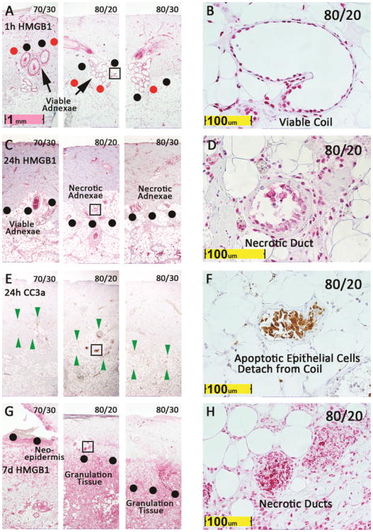
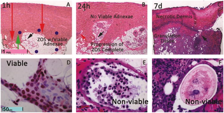
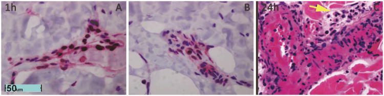
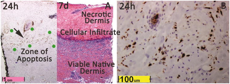
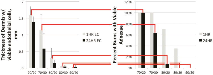
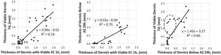
Burn injury progression has not been well characterized at the cellular level. To define burn injury progression in terms of cell death, histopathologic spatiotemporal relationships of cellular necrosis and apoptosis were investigated in a validated porcine model of vertical burn injury progression. Cell necrosis was identified by high mobility group box 1 protein and apoptosis by Caspase 3a staining of tissue samples taken 1 hour, 24 hours, and 7 days postburn. Level of endothelial cell necrosis at 1 hour was predictive of level of apoptosis at 24 hours (Pearson's r = 0.87) and of level of tissue necrosis at 7 days (Pearson's r = 0.87). Furthermore, endothelial cell necrosis was deeper than interstitial cell necrosis at 1 hour (p < 0.001). Endothelial cell necrosis at 1 hour divided the zone of injury progression (Jackson's zone of stasis) into an upper subzone with necrotic endothelial cells and initially viable adnexal and interstitial cells at 1 hour that progressed to necrosis by 24 hours and a lower zone with initially viable endothelial cells at 1 hour but necrosis and apoptosis of all cell types by 24 hours. Importantly, this spatiotemporal series of events and rapid progression resembles myocardial infarction and stroke and implicates mechanisms of these injuries, ischemia, ischemia reperfusion, and programmed cell death in burn progression.
© 2013 by the Wound Healing Society.
Conflict of interest statement
Figures
Similar articles
-
Spatiotemporal progression of cell death in the zone of ischemia surrounding burns.Wound Repair Regen. 2011 Sep-Oct;19(5):622-32. doi: 10.1111/j.1524-475X.2011.00725.x. Wound Repair Regen. 2011. PMID: 22092800 Free PMC article.
-
Histopathologic staining of low temperature cutaneous burns: comparing biomarkers of epithelial and vascular injury reveals utility of HMGB1 and hematoxylin phloxine saffron.Wound Repair Regen. 2012 Nov-Dec;20(6):918-27. doi: 10.1111/j.1524-475X.2012.00847.x. Wound Repair Regen. 2012. PMID: 23126459
-
Apoptosis and necrosis in the ischemic zone adjacent to third degree burns.Acad Emerg Med. 2008 Jun;15(6):549-54. doi: 10.1111/j.1553-2712.2008.00115.x. Acad Emerg Med. 2008. PMID: 18616442
-
A review of the local pathophysiologic bases of burn wound progression.J Burn Care Res. 2010 Nov-Dec;31(6):849-73. doi: 10.1097/BCR.0b013e3181f93571. J Burn Care Res. 2010. PMID: 21105319 Review.
-
Research advances in prevention and treatment of burn wound deepening in early stage.Front Surg. 2022 Oct 21;9:1015411. doi: 10.3389/fsurg.2022.1015411. eCollection 2022. Front Surg. 2022. PMID: 36338639 Free PMC article. Review.
Cited by
-
Current concepts on burn wound conversion-A review of recent advances in understanding the secondary progressions of burns.Burns. 2016 Aug;42(5):1025-1035. doi: 10.1016/j.burns.2015.11.007. Epub 2016 Jan 17. Burns. 2016. PMID: 26787127 Free PMC article. Review.
-
Metal chelation reduces skin epithelial inflammation and rescues epithelial cells from toxicity due to thermal injury in a rat model.Burns Trauma. 2020 Oct 2;8:tkaa024. doi: 10.1093/burnst/tkaa024. eCollection 2020. Burns Trauma. 2020. PMID: 33033727 Free PMC article.
-
Utility of spatial frequency domain imaging (SFDI) and laser speckle imaging (LSI) to non-invasively diagnose burn depth in a porcine model.Burns. 2015 Sep;41(6):1242-52. doi: 10.1016/j.burns.2015.03.001. Epub 2015 Jun 30. Burns. 2015. PMID: 26138371 Free PMC article.
-
It Is Written in the Clot: Coagulation Assessment in Severe Burn Injury.Eur Burn J. 2025 Jun 24;6(3):37. doi: 10.3390/ebj6030037. Eur Burn J. 2025. PMID: 40700333 Free PMC article.
-
Thermal injury induces early blood vessel occlusion in a porcine model of brass comb burn.Sci Rep. 2021 Jun 14;11(1):12457. doi: 10.1038/s41598-021-91874-0. Sci Rep. 2021. PMID: 34127701 Free PMC article.
References
-
- Singh V, Devgan L, Bhat S, Milner SM. The pathogenesis of burn wound conversion. Ann Plast Surg. 2007;59(1):109–15. - PubMed
-
- Jackson DM. Diagnosis of the Depth of Burning. Br J Surg. 1953:588–596. - PubMed
-
- Langton AK, Herrick SE, Headon DJ. An extended epidermal response heals cutaneous wounds in the absence of a hair follicle stem cell contribution. J Invest Dermatol. 2008;128:1311–1318. - PubMed
-
- Jackson DM. Second Thoughts on the Burn Wound. J Trauma. 1969;9(10):839–62. - PubMed
-
- Shupp JW, Nasabzadeh TJ, Rosenthal DS, Jordan MH, Filder P, Jeng JC. A Review of the Local Pathophysiologic Bases of Burn Wound Progression. J Burn Care Res. 2010;31(6):849–73. - PubMed
Publication types
MeSH terms
Substances
Grants and funding
LinkOut - more resources
Full Text Sources
Other Literature Sources
Medical
