. 2013 Apr;19(4):534-41.
doi: 10.3201/eid1904.121390.

Circovirus in tissues of dogs with vasculitis and hemorrhage

Affiliations

Circovirus in tissues of dogs with vasculitis and hemorrhage

Linlin Li et al. Emerg Infect Dis. 2013 Apr.

Abstract

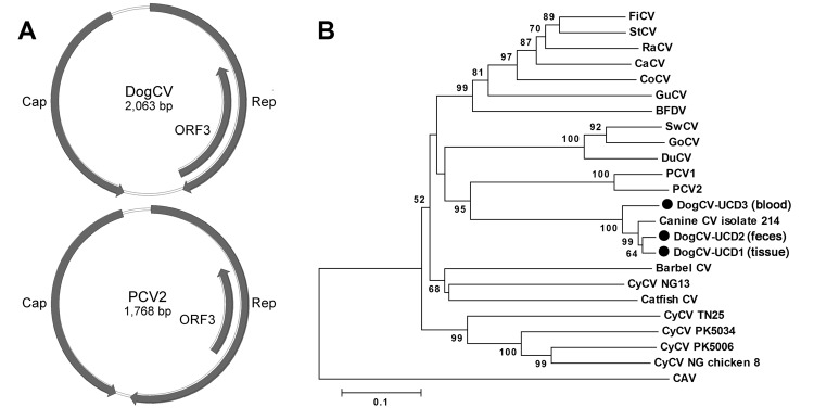
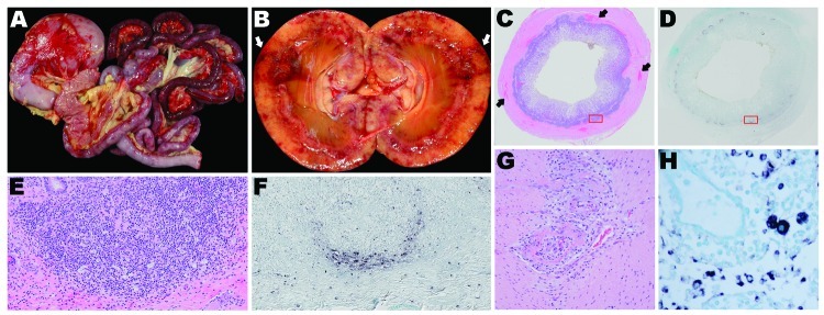
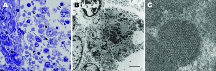
We characterized the complete genome of a novel dog circovirus (DogCV) from the liver of a dog with severe hemorrhagic gastroenteritis, vasculitis, and granulomatous lymphadenitis. DogCV was detected by PCR in fecal samples from 19/168 (11.3%) dogs with diarrhea and 14/204 (6.9%) healthy dogs and in blood from 19/409 (3.3%) of dogs with thrombocytopenia and neutropenia, fever of unknown origin, or past tick bite. Co-infection with other canine pathogens was detected for 13/19 (68%) DogCV-positive dogs with diarrhea. DogCV capsid proteins from different dogs varied by up to 8%. In situ hybridization and transmission electron microscopy detected DogCV in the lymph nodes and spleens of 4 dogs with vascular compromise and histiocytic inflammation. The detection of a circovirus in tissues of dogs expands the known tropism of these viruses to a second mammalian host. Our results indicate that circovirus, alone or in co-infection with other pathogens, might contribute to illness and death in dogs.

Keywords: canine vascular disease; circovirus; deep sequencing; dogs; granulomatous lymphadenitis; hemorrhagic gastroenteritis; necrotizing vasculitis; vasculitis; viruses.


Figures

Figure 1
Figure 1
A) Genome organization of dog circovirus (DogCV) and porcine circovirus 2 (PCV2). B) Phylogenetic analysis of DogCV strains (UCD1–3, isolated from tissue, feces, and blood respectively) based on the amino acid sequence of the replicate (Rep) protein. GenBank accession numbers for circoviruses used in the analysis: Finch circovirus (FiCV), DQ845075; Starling circovirus (StCV), DQ172906; Raven circovirus (RaCV), DQ146997; canary circovirus (CaCV), AJ301633); Columbid circovirus (CoCV), AF252610; Gull circovirus (GuCV), DQ845074; beak and feather disease virus (BFDV), AF071878; Cygnus olor circovirus (SwCV), EU056310; Goose circovirus (GoCV), AJ304456; Duck circovirus (DuCV), DQ100076; Porcine circovirus 1 (PCV1), AY660574; PCV2, AY424401; canine CV, JQ821392; Barbel CV, GU799606; Cyclovirus (CyCV) NG13, GQ404856; Silurus glanis circovirus (Catfish CV), JQ011378; CyCV TN25, GQ404857; CyCV PK5034, GQ404845; CyCV PK5006, GQ404844; CyCV NG Chicken8, HQ738643); and chicken anemia virus (CAV), M55918.
Figure 2
Figure 2
Organ tissues from sentinel dog (dog 1) and 2 other dogs (dogs 2 and 3) that were positive by in situ hybridization (ISH) analysis for dog circovirus (DogCV). A) Gross view of the gastrointestinal system from dog 1. Multifocal to coalescing hemorrhages are shown in the stomach and intestinal serosa. B) Gross view of the kidney from dog 2. Segmental regions of hemorrhage and necrosis (infarction) can be seen within the cortex and radiating from the medullary papilla to the capsule. C) Hemolyxin and eosin (H&E) stain of the ileum from dog 1. Peyer’s patches are moderately depleted, and multifocal regions of hemorrhage are present within the muscular intestinal wall. D) ISH of the ileum from dog 1. Peyer’s patches circumferentially contain abundant DogCV DNA. E) H&E stain of a Peyer’s patch in the ileum from dog 1, showing moderate depletion of lymphocytes and an increased population of macrophages within the germinal center and the peripheral base of the follicle. F) ISH of a Peyer’s patch in the ileum from dog 1. DogCV DNA is rich within the cytoplasm of abundant cells at the periphery of the follicle, and individual cells are scattered within the germinal center, lymphatic channels, and submucosa. The positive cells have the morphologic appearance of macrophages. G) H&E stain of the jejunum from dog 3, showing segmental, circumferential, fibrinoid necrosis of the artery. H) ISH of a mesenteric lymph node from dog 2. Macrophages in the medullary sinus and lymphatic cords contain abundant DogCV DNA.
Figure 3
Figure 3
Lymph node from sentinel dog from which dog circovirus was identified. A) Toluidine blue stain shows multiple macrophages within the medullary sinus contain vacuoles and discrete, oblong to round, variably stained cytoplasmic bodies (arrows). B) A single macrophage adjacent to a lymphocyte (upper left) and partial profiles of other cells. Intracytoplasmic inclusion bodies are distributed throughout the macrophage cytoplasm, along with mitochondria and vacuoles. Scale bar indicates 2 µm. C) Intracytoplasmic inclusion bodies contain granular content and sometimes paracrystalline to herringbone arrays of 10–11 nm diameter viral-like particles. Scale bar indicates 100 nm.

Similar articles

Cited by

References

    1. Li L, Kapoor A, Slikas B, Bamidele OS, Wang C, Shaukat S, et al. Multiple diverse circoviruses infect farm animals and are commonly found in human and chimpanzee feces. J Virol. 2010;84:1674–82. 10.1128/JVI.02109-09 - DOI - PMC - PubMed
    1. Todd D. Avian circovirus diseases: lessons for the study of PMWS. Vet Microbiol. 2004;98:169–74. 10.1016/j.vetmic.2003.10.010 - DOI - PubMed
    1. Allan G, Krakowka S, Ellis J, Charreyre C. Discovery and evolving history of two genetically related but phenotypically different viruses, porcine circoviruses 1 and 2. Virus Res. 2012;164:4–9. 10.1016/j.virusres.2011.09.013 - DOI - PubMed
    1. Segalés J. Porcine circovirus type 2 (PCV2) infections: clinical signs, pathology and laboratory diagnosis. Virus Res. 2012;164:10–9. 10.1016/j.virusres.2011.10.007 - DOI - PubMed
    1. Opriessnig T, Meng XJ, Halbur PG. Porcine circovirus type 2 associated disease: update on current terminology, clinical manifestations, pathogenesis, diagnosis, and intervention strategies. J Vet Diagn Invest. 2007;19:591–615. 10.1177/104063870701900601 - DOI - PubMed

Publication types

MeSH terms