Review
. 2013 Apr;23(2):277-84.
doi: 10.1016/j.sbi.2013.04.005. Epub 2013 Apr 26.

Unanticipated parallels in architecture and mechanism between ATP-gated P2X receptors and acid sensing ion channels

Affiliations
Review

Unanticipated parallels in architecture and mechanism between ATP-gated P2X receptors and acid sensing ion channels

Isabelle Baconguis et al. Curr Opin Struct Biol. 2013 Apr.

Abstract

ATP-gated P2X receptors and acid-sensing ion channels are cation-selective, trimeric ligand-gated ion channels unrelated in amino acid sequence. Nevertheless, initial crystal structures of the P2X4 receptor and acid-sensing ion channel 1a in resting/closed and in non conductive/desensitized conformations, respectively, revealed common elements of architecture. Recent structures of both channels have revealed the ion channels in open conformations. Here we focus on common elements of architecture, conformational change and ion permeation, emphasizing general principles of structure and mechanism in P2X receptors and in acid-sensing ion channels and showing how these two sequence-disparate families of ligand-gated ion channel harbor unexpected similarities when viewed through a structural lens.


Figures

Figure 1
Figure 1
P2X4 and ASIC1a architecture, domain organization and agonist or toxin binding site. a. Zebrafish P2X4 receptor structure in the apo, closed state, color coded by domain. b. Domain organization of a single P2X4 apo subunit. c. Domain organization of cASIC1a. d. Chicken ASIC1a structure in the low pH, desensitized, non conductive state. e. P2X4 structure in the ATP-bound, open channel state. f. cASIC1a bound to psalmotoxin (PcTx1) at low pH. g. cASIC1a bound to PcTx1 at high pH.
Figure 2
Figure 2
Binding sites for ATP and toxin in P2X4 receptor and ASIC1a, respectively, flank the central β-sheet ‘scaffold’ domains. a. Close up of the intersubunit binding site for ATP in the P2X4 receptor. b. Close up of the intersubunit binding site for PcTx1. ATP and the toxin are shown in sphere representation, color coded by atom type (carbon: gray; oxygen: red; nitrogen: blue; sulfur: yellow). The proteins are colored as in Figure 1.
Figure 3
Figure 3
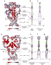
The P2X4 receptor and ASIC1a harbor cavities, vestibules and lateral fenestrations. a. Surface representation of the P2X4 receptor color coded by electrostatic potential. ‘HOLE’ representation of the apo, closed (b) and ATP-bound, open (c) states of the P2X4 receptor, showing a large pore in the ATP-bound state. d. Surface representation of ASIC1a also colored by electrostatic potential. ‘HOLE’ representations of ASIC1a in the low pH desensitized state (e), the high pH PcTx1 bound state (f) and the low pH PcTx1 bound state, illustrating a large non-selective pore in (f) and small, asymmetric and selective pore in (g). In both the P2X4 receptor and ASIC1a ions enter and exit the pore from the extracellular site of the membrane by way of lateral fenestrations. For both channels a possible ion pathway along the 3-fold axis within the extracellular domain remains occluded.
Figure 4
Figure 4
Conformational changes accompanying gating in the P2X4 receptor and ASIC1a. a. Superposition of the upper body domain – the ‘scaffold’ - of the apo (gray) and ATP-bound states of the P2X4 receptor showing how the β-strands of the lower body domain flex outward in the ATP-bound state. A superposition of the upper palm and knuckle domains – the ‘scaffold’ – of the low pH desensitized state with the high pH PcTx1 state (b) and the low pH PcTx1 state (c), illustrating how the scaffold domain is relatively immobile whereas the β-strands of the lower palm domain flex outward in the PcTx1 bound states.
Figure 5
Figure 5
Cartoon schematic illustrating the core domains involved in the ATP-dependent and PcTx1-dependent gating of the P2X4 receptor and ASIC1a, respectively. The ‘effector binding domains’ are, as examples, the ‘head’, ‘body’, ‘dorsal fin’ and ‘left flipper’ in the case of the P2X4 receptor and the ‘finger’, ‘thumb’ and ‘palm’ domains of ASIC1a.

Similar articles

Cited by

References

    1. Cowan WM, Sudhof TC, Stevens CF, editors. Synapses. The Johns Hopkins University Press; Baltimore, MD: 2001.
    1. Khakh BS, North RA. Neuromodulation by extracellular ATP and P2X receptors in the CNS. Neuron. 2012;76:51–69. - PMC - PubMed
    1. Sherwood TW, Frey EN, Askwith CC. Structure and activity of the acid-sensing ion channels. Am J Physiol Cell Physiol. 2012;303:C699–710. - PMC - PubMed
    1. Gründer S, Chen X. Structure, function, and pharmacology of acid-sensing ion channels (ASICs): focus on ASIC1a. Int J Physiol Pathophysiol Pharmacol. 2010;2:73–94. - PMC - PubMed
    1. Mayer ML. Emerging models of glutamate receptor ion channel strucure and function. Structure. 2011;19:1370–1380. - PMC - PubMed

Publication types

MeSH terms