. 2013 Jun;109(2):183-93.
doi: 10.1016/j.ymgme.2013.03.013. Epub 2013 Apr 6.

The effect of neonatal gene therapy on skeletal manifestations in mucopolysaccharidosis VII dogs after a decade

Affiliations

The effect of neonatal gene therapy on skeletal manifestations in mucopolysaccharidosis VII dogs after a decade

Elizabeth M Xing et al. Mol Genet Metab. 2013 Jun.

Abstract

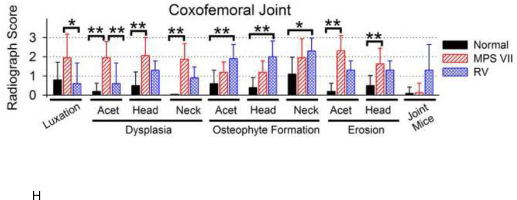
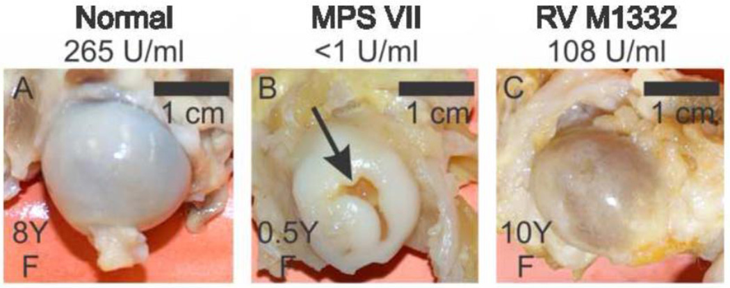
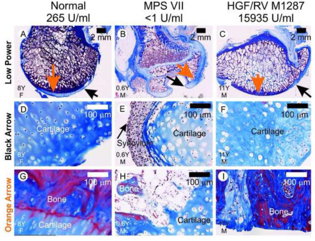
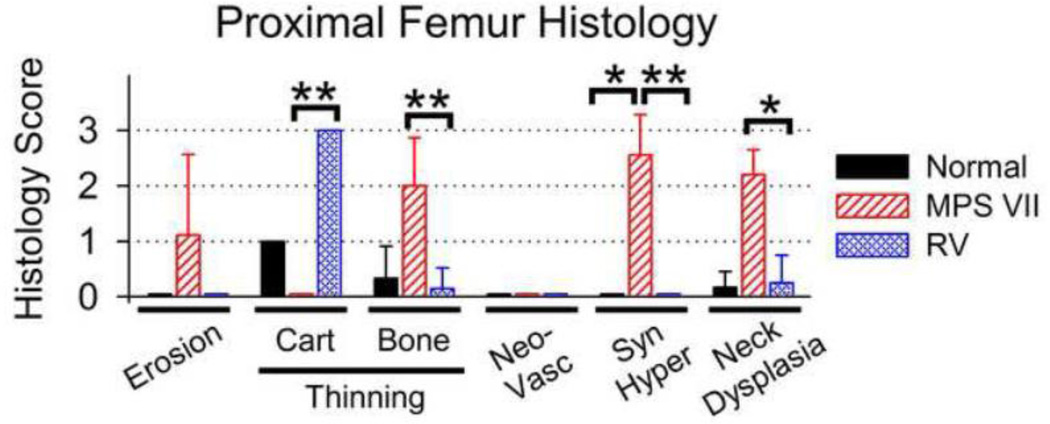
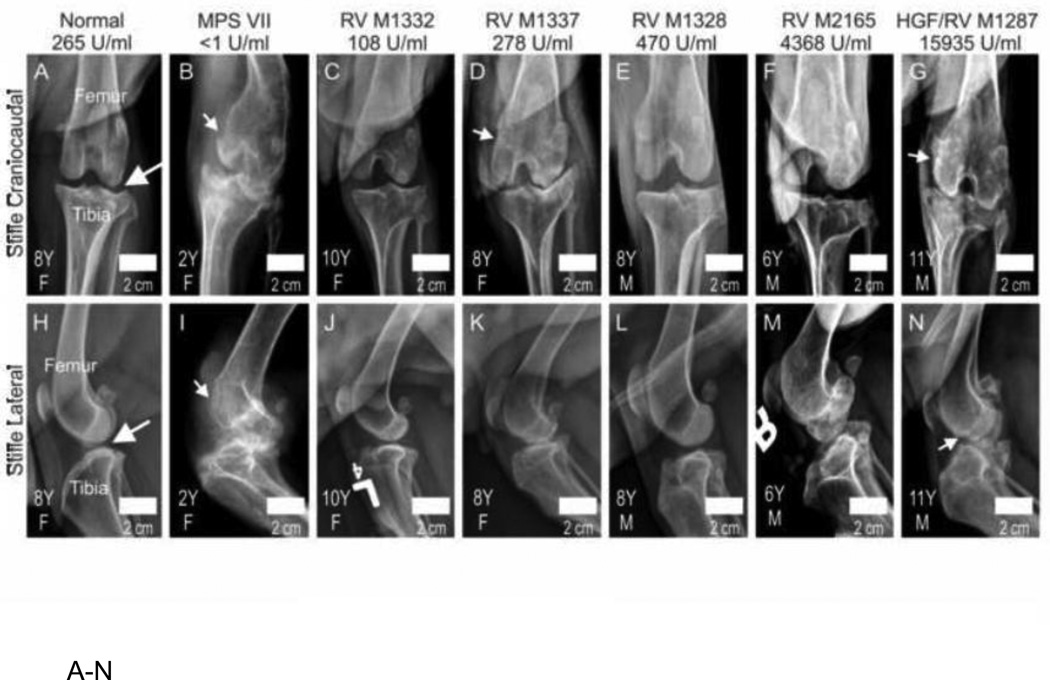
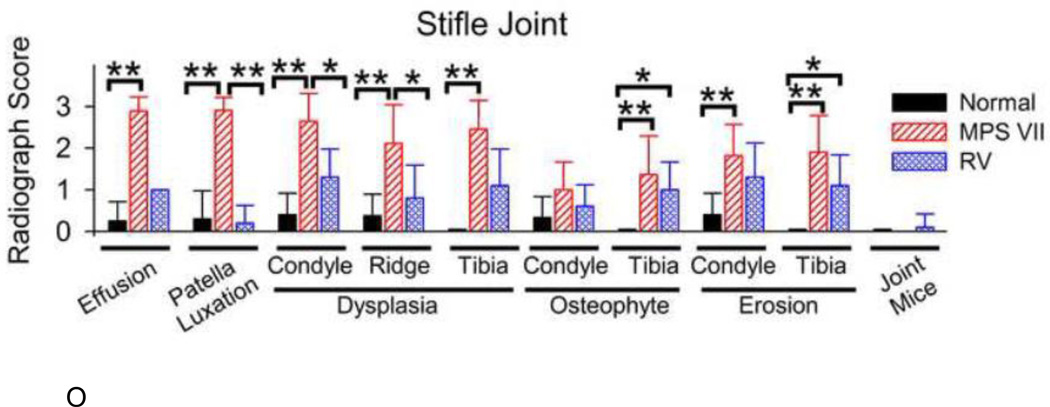
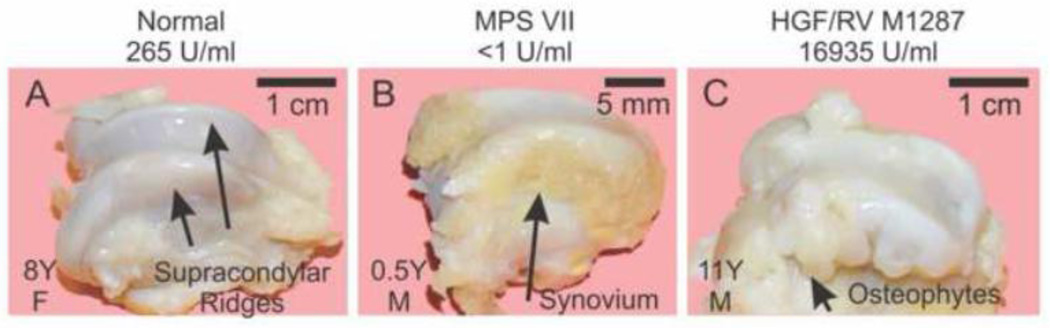
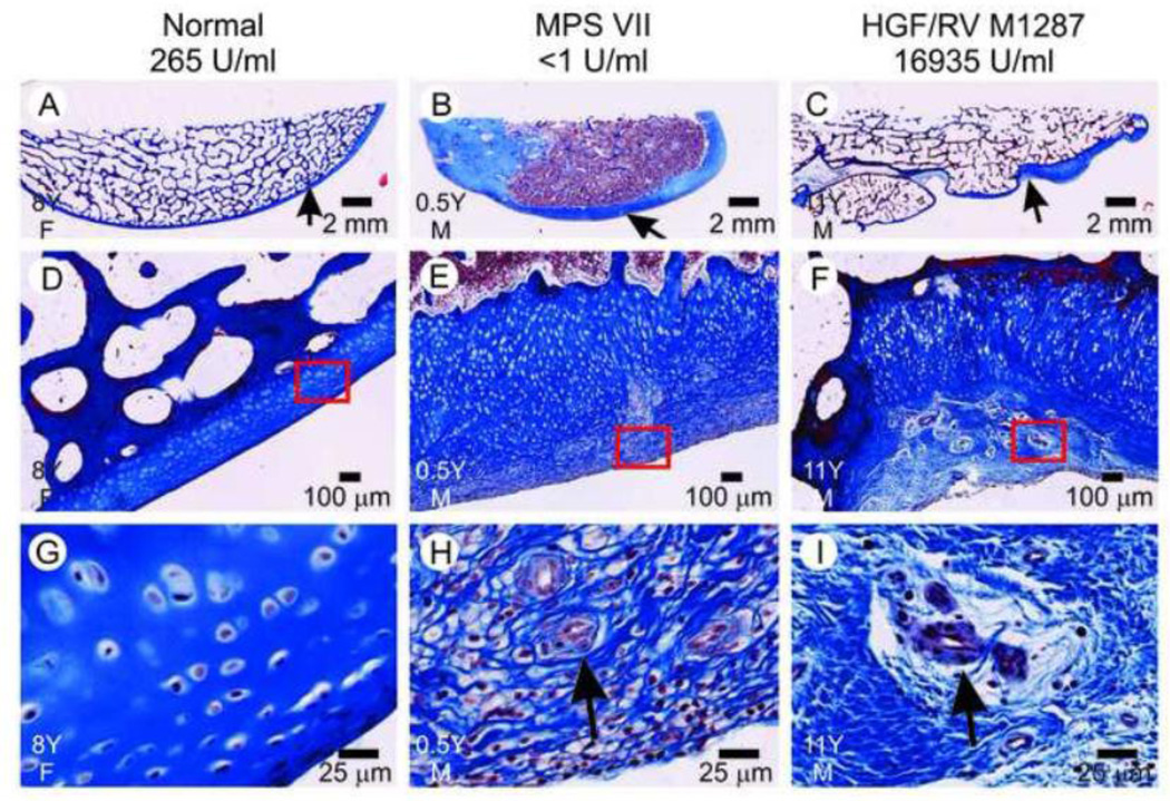
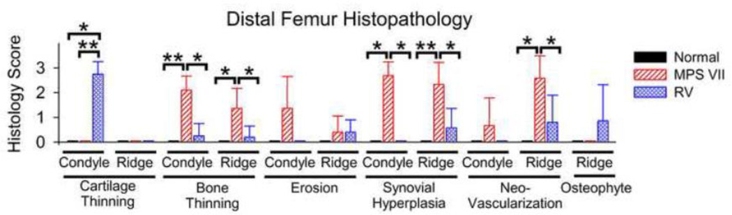
Mucopolysaccharidosis (MPS) VII is a lysosomal storage disease due to deficient activity of β-glucuronidase (GUSB), and results in glycosaminoglycan accumulation. Skeletal manifestations include bone dysplasia, degenerative joint disease, and growth retardation. One gene therapy approach for MPS VII involves neonatal intravenous injection of a gamma retroviral vector expressing GUSB, which results in stable expression in liver and secretion of enzyme into blood at levels predicted to be similar or higher to enzyme replacement therapy. The goal of this study was to evaluate the long-term effect of neonatal gene therapy on skeletal manifestations in MPS VII dogs. Treated MPS VII dogs could walk throughout their lives, while untreated MPS VII dogs could not stand beyond 6 months and were dead by 2 years. Luxation of the coxofemoral joint and the patella, dysplasia of the acetabulum and supracondylar ridge, deep erosions of the distal femur, and synovial hyperplasia were reduced, and the quality of articular bone was improved in treated dogs at 6 to 11 years of age compared with untreated MPS VII dogs at 2 years or less. However, treated dogs continued to have osteophyte formation, cartilage abnormalities, and an abnormal gait. Enzyme activity was found near synovial blood vessels, and there was 2% as much GUSB activity in synovial fluid as in serum. We conclude that neonatal gene therapy reduces skeletal abnormalities in MPS VII dogs, but clinically-relevant abnormalities remain. Enzyme replacement therapy will probably have similar limitations long-term.


Figures

Fig. 1
Fig. 1. Radiographs of coxofemoral joints in dogs
A–G. Representative radiographs. Representative craniocaudal radiographs of the coxofemoral joint are presented from 1 normal, 1 untreated MPS VII (MPS VII), and 5 gene therapy-treated MPS VII dogs as indicated above the panel. Treated MPS VII dogs received a neonatal IV injection of the retroviral vector hAAT-cGUSB-WPRE either with preceding hepatocyte growth (HGF/RV) or without preceding HGF (RV). The average lifetime serum GUSB activity for dogs is indicated, and treated dogs are arranged from lowest (left) to highest (right) serum GUSB activity. Radiographs were obtained at the indicated age in years (Y) for females (F) or males (M), as indicated in the lower left corner. The white arrow in panel A indicates the neck of the femur for a normal dog. For the MPS VII dog in panel B, the femur is luxated from the acetabulum and the head and neck are dysplastic. Size bars indicate 2 cm. H. Radiographic scores of coxofemoral joints. Radiographs were scored from 0 (normal) to +3 (severely abnormal) for luxation of the femur from the acetabulum, dysplasia, osteophyte formation, and/or surface erosion of the acetabulum (Acet), femoral head (Head) and femoral neck (Neck), and the average number of joint mice (calcified stones) per joint. Scores are shown for 10 coxofemoral joints from 5 normal dogs (age 8.1±0.7 years), 16 joints from 8 untreated MPS VII dogs (age 1.6 ±0.4 years), and 10 joints from 5 RV-treated MPS VII dogs (age 8.2±1.9 years). The mean ± standard deviation (SD) is shown; (*) indicates p<0.05, and (**) indicates p<0.01 for comparison of the groups connected by a bracket using the Kruskal-Wallis one-way analysis of variance on ranks with Dunn’s post-hoc analysis. Untreated and treated MPS VII dogs had approximately equal numbers of males and females, while normal dogs were all females from the colony that were used for breeding purposes. Three coxofemoral joints from untreated MPS VII dogs could not be evaluated for osteophyte formation or articular erosion as the dysplasia was so severe.
Fig. 2
Fig. 2. Gross evaluation of the head of the proximal femur
Post-mortem examination was performed for animals of the indicated genotype and treatment group (top) at the indicated age in years (Y) for animals of the indicated gender (M or F) (lower left corner), and photographs were obtained. The normal dog had a bright white shiny smooth surface of the head of the femur. An untreated MPS VII dog maintained a bright white shiny color over most of the head of the femur, but had a deep fissure that was 3 mm deep. The HGF/RV-treated dog did not have any erosions, but the color of the surface was less white and shiny than in a normal dog, suggesting that the amount of cartilage on the surface was reduced.
Fig. 3
Fig. 3. Histology of the proximal femurs
Formalin-fixed samples were embedded in paraffin and 6 µm-thick sections were stained with Masson’s trichrome, which stains collagen blue. The treatment group is indicated above the panel, and the age in years (Y) and the gender (M or F) are indicated in the lower left hand corner. The black arrows in panels A–C identify a region of cartilage in each dog that is shown at higher power in panels D–F. The black arrow in panel E identifies synovium on the surface of cartilage in an untreated MPS VII dog. The orange arrows in panels A–C indicate the areas at the bone:cartilage interface that are shown at higher magnification in panels G–I. For the MPS VII dog in panel H, there are regions where the bone is discontinuous under the cartilage layer. For the treated MPS VII dog in panel I, there is a loss of cartilage over most of the articular surface.
Fig. 4
Fig. 4. Scoring of histology of proximal femurs
Sections of bones that were stained with Masson’s trichrome as shown in Fig. 3 were scored for erosion of the articular surface, diffuse thinning of the cartilage (Cart) or the underlying bone (Bone) on the articular surface, neovascularization (Neovasc), synovial hyperplasia (Syn Hyper), and for dysplasia of the neck of the femur using a score from 0 (normal) to +3 (severely abnormal). The mean ± SD is shown, and statistics were performed as in Fig. 1H. The mean age for collection of samples for normal, untreated MPS VII, and treated MPS VII dogs was 8.6±0.6, 0.6±0.1, and 8.7±2.1 years, respectively.
Fig. 5
Fig. 5. Radiographs of the stifle joints in dogs
A–N. Representative radiographs. Craniocaudal (top) or lateral (bottom) radiographs of the stifle joint of dogs of the indicated genotype and treatment, age in years (Y), and gender (M or F; see lower left), were obtained as described in Fig. 1. The average lifetime serum GUSB activity for treated dogs is indicated, and average serum GUSB activity in individual dogs increases from left to right. The long white arrows in panels A and H indicate the non-calcified joint space between the femur and the tibia of a normal dog. The short white arrows in some of the other panels identify erosions of the bone. O. Scoring of radiographs. The stifle joint was scored on a scale of 0 (normal) to +3 (severely abnormal) for effusion, patellar luxation, dysplasia, osteophyte formation, and surface erosion of the condyle (Condyle), the supracondylar ridge (Ridge), and the tibia, and for joint mice. The mean score ± SD was calculated and statistical comparisons performed as in Fig. 1. The mean age for obtaining radiographs was 8.1±0.7, 1.6 ±0.4, and 8.2±1.9 years for normal, untreated MPS VII, and treated MPS VII dogs, respectively.
Fig. 6
Fig. 6. Gross photographs of the distal femur
Gross photographs were taken of the distal femur after dissection of the stifle joint of dogs of the indicated group at the indicated years (Y) of age and gender (M or F). The supracondylar ridges are identified with black arrows for a normal dog in panel A. Synovial hyperplasia resulted in the tan tissue at the side of the supracondylar ridge in the MPS VII dog in panel B (long arrow). Osteophytes are identified in the HGF/RV-treated dog in panel C.
Fig. 7
Fig. 7. Histopathology of the supracondylar ridge
The supracondylar ridge of dogs of the indicated genotype and treatment group that were of the indicated age in years (Y) and gender (M or F) was processed and stained with Masson’s trichrome. The short arrows in panels A–C indicate the area at the surface of the supracondylar ridge that is shown at higher magnification in panels D–F; the red box in the latter panels indicates a region that is shown at still higher magnification in panels G–I. The black arrows in panels H and I indicate blood vessels.
Fig. 8
Fig. 8. Scoring of histology of distal femurs
Sections of bones that were stained with Masson’s trichrome as shown in Fig. 7 were scored for diffuse thinning of the cartilage or the underlying bone, erosion of the articular surface, synovial hyperplasia, neovascularization, and for osteophyte formation of the condyles or the supracondylar ridge (Ridge) of the distal femur using a score from 0 (normal) to +3 (severely abnormal). The mean ± SD is shown, and statistics were performed as in Fig. 1H. The mean age for collection of samples for normal, untreated MPS VII, and treated MPS VII dogs was 8.6±0.6, 0.6±0.1, and 8.7±2.1 years, respectively.

Similar articles

Cited by

References

    1. Neufeld EF, Muenzer J. The Mucopolysaccharidoses. In: Scriver CR, Beaudet AL, Sly WS, Valle D, editors. The metabolic and molecular bases of inherited disease. New York: McGraw-Hill; 2001. p. 3421.
    1. Moore D, Connock MJ, Wraith E, Lavery C. The prevalence of and survival in Mucopolysaccharidosis I: Hurler, Hurler-Scheie and Scheie syndromes in the UK. Orphanet J Rare Dis. 2008 Sep;16(3):24. - PMC - PubMed
    1. White KK, Harmatz P. Orthopedic management of mucopolysaccharide disease. J Pediatr Rehabil Med. 2010;3:47–56. - PubMed
    1. White KK. Orthopaedic aspects of mucopolysaccharidoses. Rheumatology (Oxford) 2011;50(Suppl 5):v26–V33. - PubMed
    1. Masterson EL, Murphy PG, O'Meara A, Moore DP, Dowling FE, Forgarty EE. Hip dysplasia in Hurler's syndrome: orthopaedic management after bone marrow transplantation. J Pediatr Orthop. 1996;16:731–733. - PubMed

Publication types

LinkOut - more resources