. 2013 Jun;23(6):835-50.
doi: 10.1038/cr.2013.59. Epub 2013 Apr 30.

Dlic1 deficiency impairs ciliogenesis of photoreceptors by destabilizing dynein

Affiliations

Dlic1 deficiency impairs ciliogenesis of photoreceptors by destabilizing dynein

Shanshan Kong et al. Cell Res. 2013 Jun.

Erratum in

  • Cell Res. 2013 Jul;23(7):972. Du, Xinrong [corrected to Du, Xingrong]

Abstract

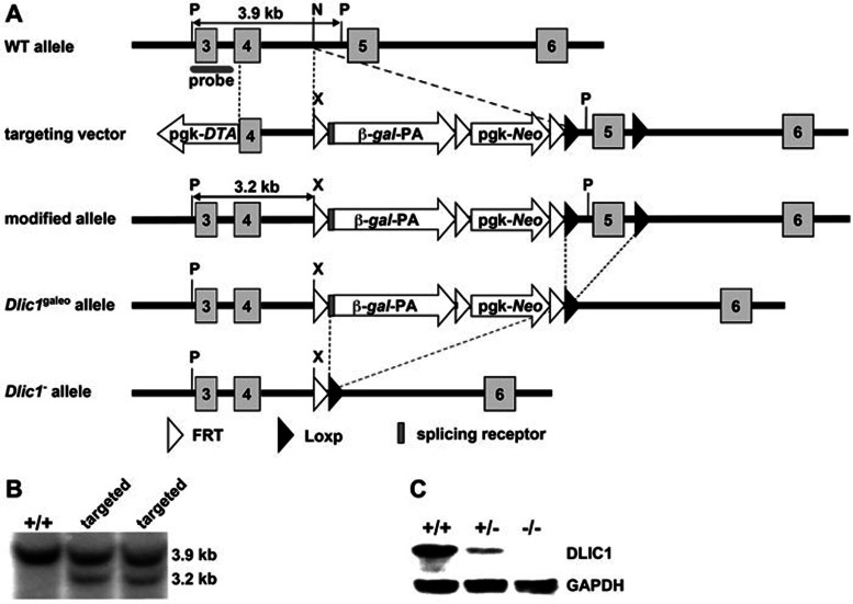
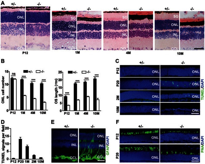
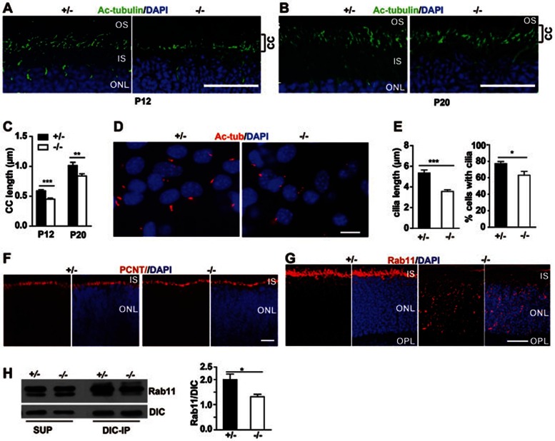
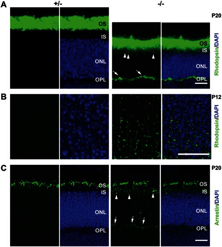
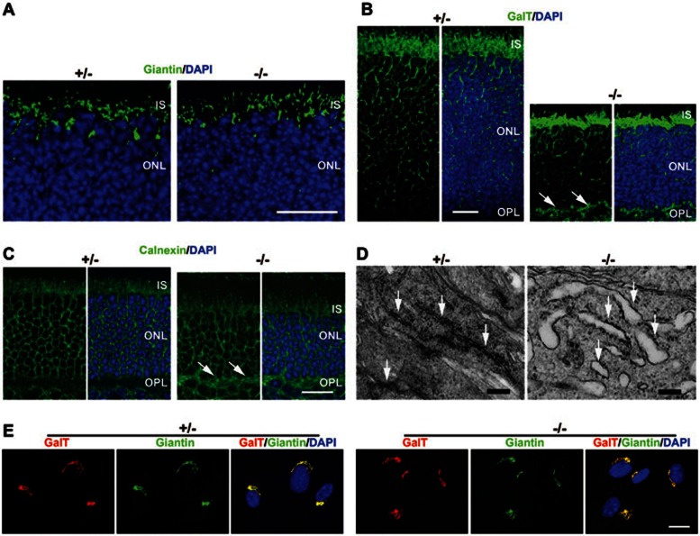
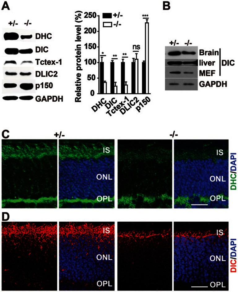
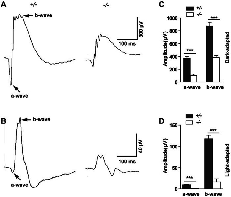
Cytoplasmic dynein 1 is fundamentally important for transporting a variety of essential cargoes along microtubules within eukaryotic cells. However, in mammals, few mutants are available for studying the effects of defects in dynein-controlled processes in the context of the whole organism. Here, we deleted mouse Dlic1 gene encoding DLIC1, a subunit of the dynein complex. Dlic1(-/-) mice are viable, but display severe photoreceptor degeneration. Ablation of Dlic1 results in ectopic accumulation of outer segment (OS) proteins, and impairs OS growth and ciliogenesis of photoreceptors by interfering with Rab11-vesicle trafficking and blocking efficient OS protein transport from Golgi to the basal body. Our studies show that Dlic1 deficiency partially blocks vesicle export from endoplasmic reticulum (ER), but seems not to affect vesicle transport from the ER to Golgi. Further mechanistic study reveals that lack of Dlic1 destabilizes dynein subunits and alters the normal subcellular distribution of dynein in photoreceptors, probably due to the impaired transport function of dynein. Our results demonstrate that Dlic1 plays important roles in ciliogenesis and protein transport to the OS, and is required for photoreceptor development and survival. The Dlic1(-/-) mice also provide a new mouse model to study human retinal degeneration.


Figures

Figure 1
Figure 1
The generation of Dlic1 knockout mice. (A) Schematic strategy to generate Dlic1−/− mice. WT allele: genomic DNA fragment of the WT Dlic1 gene containing exons 3-6. Targeting vector: schematic structure of the Dlic1 targeting vector. Modified allele: genomic structure of the Dlic1 modified allele after homologous recombination. Dlic1galeo allele: the Dlic1 null allele carrying the β-gal reporter fused in frame to exon 4 of Dlic1 after removing a fragment containing exon 5 by CRE. Dlic1 allele: the Dlic1 null allele derived from the Dlic1galeo allele by removing β-gal and Neo expression cassette by FLP. pgk-DTA and pgk-Neo represent the diphtheria toxin A and the neomycin expression cassettes, respectively. β-gal represents a modified E. coli β-galactosidase gene containing a nuclear localization signal at its N-terminus. The arrows indicate the transcriptional direction of DTA,β-gal and Neo genes. Exons are numbered and depicted by grey boxes. FRT and loxP sequences are represented by white and black arrowheads, respectively. P, N and X indicate the PuvII, NheI and XhoI restriction sites, respectively (Note: Puv II and Nhe I are not unique sites). The gray ellipse under exon 3 in the WT allele depicts the probe used for Southern blot analysis. The expected sizes of the restriction fragments hybridized with the probe are indicated in WT and modified alleles. (B) Southern blot analysis of genomic DNA extracted from WT and targeted ES cell clones and digested with Pvu II and Xho I restriction enzymes. The 3.9 kb and 3.2 kb fragments represent WT and modified alleles, respectively. (C) Western blot analysis of the DLIC1 protein expression in mouse brains using anti-DLIC1antibody. GAPDH was used as loading control. +/+, WT; +/−, Dlic1 heterozygote; −/−, Dlic1 homozygote.
Figure 2
Figure 2
Photoreceptor degeneration and impaired OS development in the Dlic1−/− mouse retina. (A) Images of HE-stained cryosections of Dlic1+/− and Dlic1−/− mouse retinas at the indicated ages. (B) Quantitative analysis of the thickness of the ONL (left) and length of the OS (right) in Dlic1+/− and Dlic1−/− mouse retinas at the indicated ages. (C, D) Representative images of TUNEL assay (C) and quantitative analysis (D) of apoptotic cells in the ONL of Dlic1+/− and Dlic1−/− retinas at the indicated ages. (E) Representative fluorescent images of cryosections of P20 Dlic1+/− and Dlic1−/− retinas stained with anti-GFAP show upregulation of GFAP in Dlic1−/− Müller glia. (F) Representative images of PNA-stained cone cells in Dlic1+/− and Dlic1−/− retinas at P12 and P20 show the reduced number and impaired OS development of Dlic1−/− cone cells. Cell nuclei in C, E and F were stained with DAPI. Images are representative retina sections from at least three mice per group. SG = OS + IS; ONL, outer nuclear layer; INL, inner nuclear layer; GCL, ganglion cell layer; M, month; +/−, Dlic1+/−; −/−, Dlic1−/−. The values in B and D represent the means ± SEM of three mice. ns, not significant; ***P < 0.001. Bar = 50 μm.
Figure 3
Figure 3
Dlic1 deletion impairs primary ciliogenesis by disrupting Rab11-vesicle transport. (A, B) Impaired development of the connecting cilium in the Dlic1−/− photoreceptors. Cryosections of the Dlic1+/− and Dlic1−/− retinas at age of P12 (A) and P20 (B) were immunofluorescent stained with anti-Ac-tubulin. (C) Statistical analysis of the length of the connecting cilia in Dlic1+/− and Dlic1−/− mouse photoreceptors at the indicated ages. Length of cilia in five random areas per section were measured and retinas from three mice in each group were used for the analysis. (D, E) Impaired ciliogenesis in Dlic1−/− MEFs. Primary ciliogenesis of Dlic1+/− and Dlic1−/− MEFs was induced by serum starvation. Images of primary cilia stained with anti-Ac-tubulin (D) and statistical analysis of the cilia length (E, left panel) and the percentage of cells with cilia (E, right panel) in serum-starved Dlic1+/− and Dlic1−/− MEF cells (n = 32 and 44 for the control and mutant respectively). (F) Dlic1 deletion does not affect the distribution pattern of PCNT in photoreceptor cells. Cryosections of P5 Dlic1+/− and Dlic1−/− retinas were IF stained with anti-PCNT. (G) IF stained cryosections of P12 Dlic1+/− and Dlic1−/− retinas with anti-Rab11 show the misaccumulation of Rab11 vesicles in the ONL of Dlic1−/− retina. (H) IP-western blot (left pane) and quantitative analyses (right panel) show that loss of Dlic1 leads to a decreased interaction between Rab11 and dynein. Cell nuclei are stained with DAPI. CC, connecting cilium. Images are representative retina sections from at least three mice per group. The values in C, E, H represent the means ± SEM. *P < 0.05, **P < 0.01, ***P < 0.001. Bar = 20 μm.
Figure 4
Figure 4
Mislocalization of OS proteins in Dlic1−/− photoreceptor cells. (A) Immunofluorescent staining of cryosections of P20 Dlic1+/− and Dlic1−/− retinas with anti-rhodopsin reveals the mislocalization of rhodopsin in Dlic1−/− photoreceptor cells. (B) Higher magnification of P12 mouse retinas immunofluorescent -stained with anti-rhodopsin. Perinuclear mislocalized rhodopsin is only shown in the Dlic1−/− retina. (C) Immunofluorescent-stained cryosections of P20 Dlic1+/− and Dlic1−/− retinas with anti-arrestin. Arrows and arrowheads in A and C indicate the mislocalization of rhodopsin or arrestin in the OPL and the IS, respectively. Cell nuclei were stained with DAPI. Images are representative retina sections from at least three mice per group. Bar = 20 μm.
Figure 5
Figure 5
The effect of Dlic1 deficiency on the ER-to-Golgi transport in photoreceptors. (A-C) Dlic1 deficiency does not affect the Golgi structure and distribution, but leads to the mislocalization of GalT and calnexin in the OPL. Cryosections of P20 Dlic1+/− and Dlic1−/− retinas were stained with anti-giantin (A), anti-GalT (B) and anti-calnexin (C) separately. Arrows in B and C indicate the mislocalization of GalT and calnexin in the OPL, respectively. (D) Electron micrographs of photoreceptors in P20 Dlic1+/− and Dlic1−/− mice, showing the ER in the IS. Arrows in the left and right panels indicate normal or dilated ER, respectively. (E) Co-immunofluorescent staining of Dlic1+/− and Dlic1−/− MEFs with anti-GalT and anti-Giantin shows the colocalization of GalT and Giantin signals in both mutant and control MEFs. Images are representative retina sections or MEFs from at least three mice per group. Bar = 20 μm (A-C, E); 250 nm (D).
Figure 6
Figure 6
Deletion of Dlic1 disrupts the distribution of dynein in mouse retinas and destabilizes other subunits of dynein. (A) Ablation of Dlic1 leads to the decreased protein levels of dynein subunits in retinal cells. Western blot (left panel) and quantitative (right panel) analyses of the protein levels of dynein subunits in retinal cells. (B) The reduction of DIC level in Dlic1−/− brain, liver and MEFs. Cell lysates from Dlic1+/− and Dlic1−/− retinas (A) or other tissues/cells as shown (B) were immunobloted with the indicated antibodies. GAPDH was used as a loading control. (C, D) Loss of Dlic1 results in the disruption of distribution of DHC and DIC in mouse retinas. Cryosections of P12 Dlic1+/− and Dlic1−/− retinas were immunofluorescent stained with anti-DHC (C) and anti-DIC (D). Cell nuclei were stained with DAPI. Images and blots are representative from at least three mice per group. The values in A represent the means ± SEM of three mice. ns, not significant; *P < 0.05, **P < 0.01, ***P < 0.001. Bar = 20 μm.
Figure 7
Figure 7
Functional characteristics of Dlic1+/− and Dlic1−/− retinas. (A, B) Saturating ERG responses of the retinas of the mice as indicated to 510 nm flashes at an intensity of -35 log scot. cd. s/m2 under scotopic (A) and photopic (B) conditions. Each trace is the average of individual records from five mice. (C, D) Statistical analyses of the saturating amplitude of a- and b-waves of the mice as indicated under scotopic (C) and photopic (D) conditions. The Dlic1+/− and Dlic1−/− mice were 10-month-old littermates. Values in C and D represent the means ± SEM of five mice. ***P < 0.001.

Similar articles

Cited by

References

    1. Young RW. The renewal of photoreceptor cell outer segments. J Cell Biol. 1967;33:61–72. - PMC - PubMed
    1. Tai AW, Chuang JZ, Bode C, Wolfrum U, Sung CH. Rhodopsin's carboxy-terminal cytoplasmic tail acts as a membrane receptor for cytoplasmic dynein by binding to the dynein light chain Tctex-1. Cell. 1999;97:877–887. - PubMed
    1. Wolfrum U, Schmitt A. Rhodopsin transport in the membrane of the connecting cilium of mammalian photoreceptor cells. Cell Motil Cytoskeleton. 2000;46:95–107. - PubMed
    1. Ramamurthy V, Cayouette M. Development and disease of the photoreceptor cilium. Clin Genet. 2009;76:137–145. - PubMed
    1. Vallee RB, Williams JC, Varma D, Barnhart LE. Dynein: An ancient motor protein involved in multiple modes of transport. J Neurobiol. 2004;58:189–200. - PubMed

Publication types

LinkOut - more resources