. 2013 Jun;14(6):545-52.
doi: 10.1038/embor.2013.56. Epub 2013 Apr 30.

Charcot-Marie-Tooth disease-associated mutants of GDAP1 dissociate its roles in peroxisomal and mitochondrial fission

Affiliations

Charcot-Marie-Tooth disease-associated mutants of GDAP1 dissociate its roles in peroxisomal and mitochondrial fission

Nina Huber et al. EMBO Rep. 2013 Jun.

Abstract

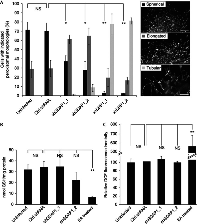
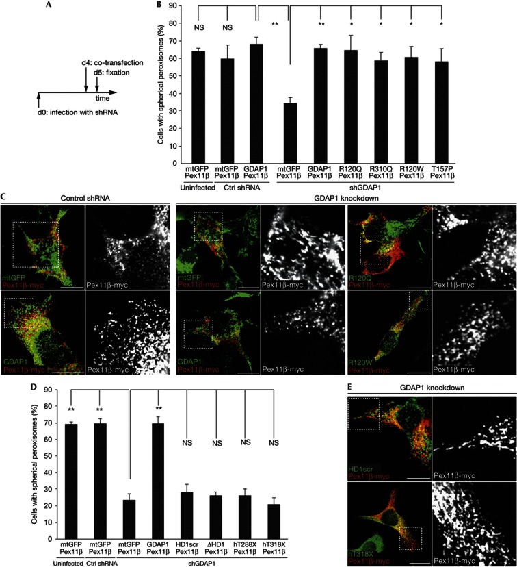
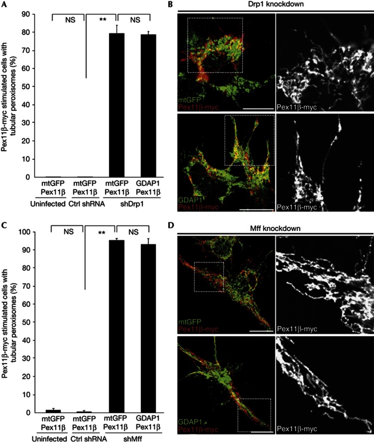
Mitochondria and peroxisomes can be fragmented by the process of fission. The fission machineries of both organelles share a set of proteins. GDAP1 is a tail-anchored protein of mitochondria and induces mitochondrial fragmentation. Mutations in GDAP1 lead to Charcot-Marie-Tooth disease (CMT), an inherited peripheral neuropathy, and affect mitochondrial dynamics. Here, we show that GDAP1 is also targeted to peroxisomes mediated by the import receptor Pex19. Knockdown of GDAP1 leads to peroxisomal elongation that can be rescued by re-expressing GDAP1 and by missense mutated forms found in CMT patients. GDAP1-induced peroxisomal fission is dependent on the integrity of its hydrophobic domain 1, and on Drp1 and Mff, as is mitochondrial fission. Thus, GDAP1 regulates mitochondrial and peroxisomal fission by a similar mechanism. However, our results reveal also a more critical role of the amino-terminal GDAP1 domains, carrying most CMT-causing mutations, in the regulation of mitochondrial compared to peroxisomal fission.


Conflict of interest statement

The authors declare that they have no conflict of interest.

Figures

Figure 1
Figure 1
GDAP1 is targeted to peroxisomes. (A) COS7 cells stably expressing GFP–SKL were transfected with GDAP1 expression constructs and stained for GDAP1 and cytochrome c. GDAP1 is mainly localized to mitochondria. Higher magnification (zoom box) shows GFP–SKL-positive structures that are positive for GDAP1 but negative for cytochrome c (arrows). A neighbouring, untransfected cell (light yellow broken line). (B) Endogenously GDAP1-expressing primary hippocampal cell cultures were infected with lentivirus encoding GFP–SKL, and stained for GDAP1 and cytochrome c. Higher magnification (zoom box) shows that endogenous GDAP1 predominantly co-localizes with the cytochrome c, and also with GFP–SKL-positive structures, which are negative for cytochrome c (arrows). (C) Measured GDAP1 fluorescence intensity in the peroxisomal population was decreased in Pex19 knockdown cells but unaffected in the mitochondrial population. Values represent mean and s.e.m. of three independent experiments. Fluorescence intensity of the peroxisomal/mitochondrial population was analysed for 6–9 cells per condition per experiment: *P<0.05, two-tailed unpaired t-test. Scale bar, 10 μm. ctrl shRNA, non-targeting shRNA control; GDAP1, ganglioside-induced differentiation associated protein 1; GFP–SKL, green fluorescent protein with peroxisomal targeting sequence; shPex19, shRNA against Pex19.
Figure 2
Figure 2
Loss of GDAP1 leads to peroxisomal elongation. (A) N1E-115 cells were infected with lentiviruses encoding shRNA against GDAP1, Drp1 or a non-targeting control (ctrl) shRNA, or were left uninfected. Five days after infection, peroxisomal morphologies were assessed after immunostaining for Pex14 according to the three categories: spherical, elongated or tubular and were quantified in blinded countings. (B) Measurements of cellular GSH levels and (C) ROS levels show no significant alterations at day five of the GDAP1 knockdown compared to control cells. EA treated cells served as positive control (50 μg/ml for 2 h). Values represent mean and s.e.m. of three independent experiments. For morphology analysis, 100 cells were counted per condition per experiment: *P<0.05, **P<0.01, two-tailed unpaired t-test. Scale bar, 2.5 μm. ctrl shRNA, non-targeting shRNA control; DCF, 2,7-dichlorofluorescin; Drp1, dynamin-related protein 1; EA, ethacrynic acid; GDAP1, ganglioside-induced differentiation associated protein 1; GSH, glutathione; NS, not significant; ROS, reactive oxygen species; s.e.m., standard error of the mean; shDrp1, shRNA against Drp1 (dynamin related protein 1); shGDAP1, shRNA against GDAP1; shRNA, short hairpin RNA.
Figure 3
Figure 3
Peroxisomal fission is dependent on the GDAP1-targeting domain and the HD1, but unaffected by CMT-disease associated N-terminal missense mutations. (A) N1E-115 cells were infected with lentiviruses encoding shRNA against GDAP1 or a non-targeting control (ctrl) shRNA or left uninfected (d0). After 4 days (d4), cells were co-transfected with Pex11β–myc and GDAP1 wildtype, mutant forms of GDAP1, or mtGFP. Twenty-four hours later (d5), cells were fixed and stained (C,E). Peroxisomal morphology of double-positive cells was quantified in a blinded counting (B,D). (GDAP1 with scrambled HD1 (HD1 scr), GDAP1 with deleted HD1 (ΔHD1), C-terminal truncated human GDAP1 (hT288X, hT318X)). Values represent means and s.e.m. of at least three independent experiments, 100 cells were counted per condition per experiment: *P<0.05, **P<0.01, two-tailed unpaired t-test. Scale bar, 10 μm. CMT, Charcot-Marie-Tooth disease; ctrl shRNA, non-targeting shRNA control; GDAP1, ganglioside-induced differentiation associated protein 1; HD1, hydrophobic domain 1; mtGFP, mitochondria-localized green fluorescent protein; NS, not significant; s.e.m., standard error of the mean; shGDAP1, shRNA against GDAP1; shRNA, short hairpin RNA.
Figure 4
Figure 4
GDAP1-induced peroxisomal fragmentation depends on Drp1 and Mff. The experiment was performed as illustrated in Fig 3A. Knockdown of Drp1 (A,B) or Mff (C,D) in combination with co-expression of Pex11β–myc and GDAP1 or mtGFP result in tubular peroxisomes. Values represent means and s.e.m. of three independent experiments, 100 cells were counted per condition per experiment: **P<0.01, two-tailed unpaired t-test. Scale bar, 10 μm. Ctrl shRNA, non-targeting shRNA control; Drp1, dynamin-related protein 1; GDAP1, ganglioside-induced differentiation associated protein 1; Mff, mitochondrial fission factor; mtGFP, mitochondria-localized green fluorescent protein; s.e.m., standard error of the mean; shMff, shRNA against Mff (mitochondrial fission factor); shRNA, short hairpin RNA.

References

    1. Wanders RJ, Waterham HR (2006) Biochemistry of mammalian peroxisomes revisited. Annu Rev Biochem 75: 295–332 - PubMed
    1. Fagarasanu A, Fagarasanu M, Rachubinski RA (2007) Maintaining peroxisome populations: a story of division and inheritance. Annu Rev Cell Dev Biol 23: 321–344 - PubMed
    1. Kim PK, Mullen RT, Schumann U, Lippincott-Schwartz J (2006) The origin and maintenance of mammalian peroxisomes involves a de novo PEX16-dependent pathway from the ER. J Cell Biol 173: 521–532 - PMC - PubMed
    1. Schrader M, Fahimi HD (2006) Growth and division of peroxisomes. Int Rev Cytol 255: 237–290 - PubMed
    1. van der Zand A, Gent J, Braakman I, Tabak HF (2012) Biochemically distinct vesicles from the endoplasmic reticulum fuse to form peroxisomes. Cell 149: 397–409 - PubMed

Publication types

MeSH terms