

Quantitative comparison of volume maintenance between inlay and onlay bone grafts in the craniofacial skeleton
- PMID: 23629083
- PMCID: PMC4487985
- DOI: 10.1097/PRS.0b013e31828e217a
Quantitative comparison of volume maintenance between inlay and onlay bone grafts in the craniofacial skeleton
Abstract
Background: Nonvascularized autologous bone grafts are the criterion standard in craniofacial reconstruction for bony defects involving the craniofacial skeleton. The authors have previously demonstrated that graft microarchitecture is the major determinant of volume maintenance for both inlay and onlay bone grafts following transplantation. This study performs a head-to-head quantitative analysis of volume maintenance between inlay and onlay bone grafts in the craniofacial skeleton using a rabbit model to comparatively determine their resorptive kinetics over time.
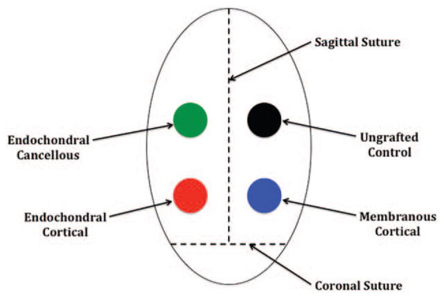
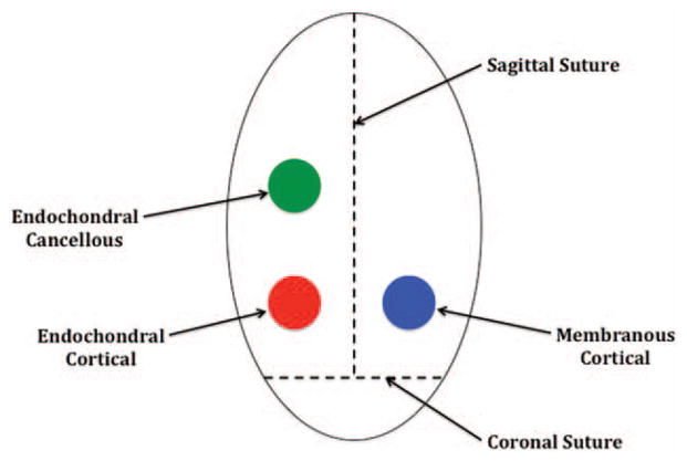
Methods: Fifty rabbits were divided randomly into six experimental groups: 3-week inlay, 3-week onlay, 8-week inlay, 8-week onlay, 16-week inlay, and 16-week onlay. Cortical bone from the lateral mandible and both cortical and cancellous bone from the ilium were harvested from each animal and placed either in or on the cranium. All bone grafts underwent micro-computed tomographic analysis at 3, 8, and 16 weeks.
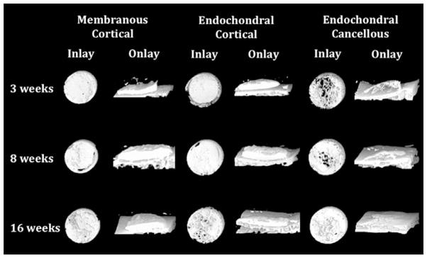
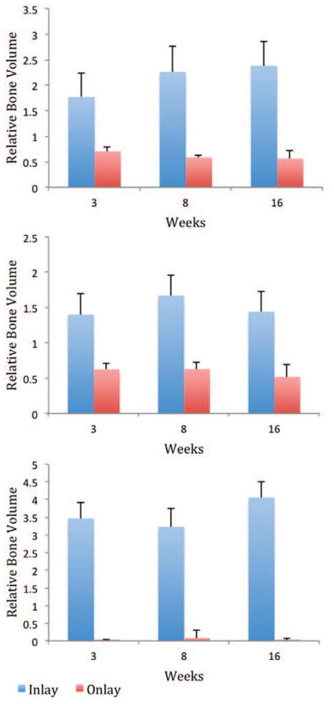
Results: All bone graft types in the inlay position increased their volume over time, with the greatest increase in endochondral cancellous bone. All bone graft types in the onlay position decreased their volume over time, with the greatest decrease in endochondral cancellous bone. Inlay bone grafts demonstrated increased volume compared with onlay bone grafts of identical embryologic origin and microarchitecture at all time points (p < 0.05).
Conclusions: Inlay bone grafts, irrespective of their embryologic origin, consistently display less resorption over time compared with onlay bone grafts in the craniofacial skeleton. Both inlay and onlay bone grafts are driven by the local mechanical environment to recapitulate the recipient bed.
Conflict of interest statement
Figures
Comment in
-
Anatomic resection of hepatocellular carcinoma: a step forward for the precise resection of the tumor-bearing portal territory of the liver.Ann Surg. 2015 May;261(5):e145. doi: 10.1097/SLA.0000000000000531. Ann Surg. 2015. PMID: 24513785 No abstract available.
-
Reply to the letter: "anatomic resection of hepatocellular carcinoma: a step forward for the precise resection of the tumor-bearing portal territory of the liver".Ann Surg. 2015 May;261(5):e145-6. doi: 10.1097/SLA.0000000000000695. Ann Surg. 2015. PMID: 24743627 No abstract available.
References
-
- Habal MB. Bone grafting in craniofacial surgery. Clin Plast Surg. 1994;21:349–363. - PubMed
-
- Donovan MG, Dickerson NC, Hellstein JW, Hanson LJ. Autologous calvarial and iliac onlay bone grafts in miniature swine. J Oral Maxillofac Surg. 1993;51:898–903. - PubMed
-
- Phillips JH, Rahn BA. Fixation effects on membranous and endochondral onlay bone graft revascularization and bone deposition. Plast Reconstr Surg. 1990;85:891–897. - PubMed
-
- Lin KY, Bartlett SP, Yaremchuk MJ, Fallon M, Grossman RF, Whitaker LA. The effect of rigid fixation on the survival of onlay bone grafts: An experimental study. Plast Reconstr Surg. 1990;86:449–456. - PubMed
Publication types
MeSH terms
Grants and funding
LinkOut - more resources
Full Text Sources
Other Literature Sources
Medical
