

Contractility and kinetics of human fetal and human adult skeletal muscle
- PMID: 23629510
- PMCID: PMC3832119
- DOI: 10.1113/jphysiol.2013.252650
Contractility and kinetics of human fetal and human adult skeletal muscle
Abstract
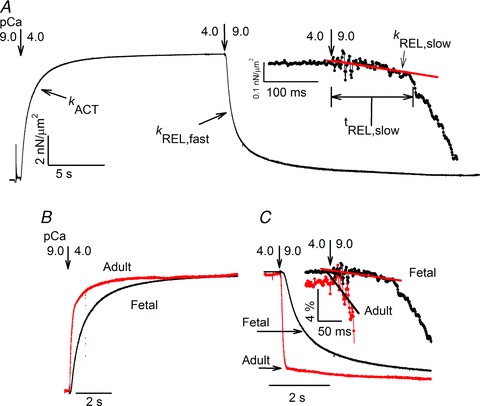
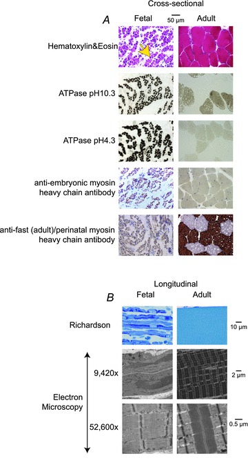
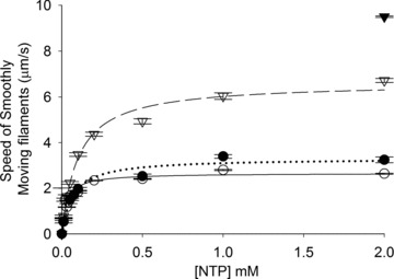
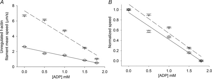
Little is known about the contraction and relaxation properties of fetal skeletal muscle, and measurements thus far have been made with non-human mammalian muscle. Data on human fetal skeletal muscle contraction are lacking, and there are no published reports on the kinetics of either fetal or adult human skeletal muscle myofibrils. Understanding the contractile properties of human fetal muscle would be valuable in understanding muscle development and a variety of muscle diseases that are associated with mutations in fetal muscle sarcomere proteins. Therefore, we characterised the contractile properties of developing human fetal skeletal muscle and compared them to adult human skeletal muscle and rabbit psoas muscle. Electron micrographs showed human fetal muscle sarcomeres are not fully formed but myofibril formation is visible. Isolated myofibril mechanical measurements revealed much lower specific force, and slower rates of isometric force development, slow phase relaxation, and fast phase relaxation in human fetal when compared to human adult skeletal muscle. The duration of slow phase relaxation was also significantly longer compared to both adult groups, but was similarly affected by elevated ADP. F-actin sliding on human fetal skeletal myosin coated surfaces in in vitro motility (IVM) assays was much slower compared with adult rabbit skeletal myosin, though the Km(app) (apparent (fitted) Michaelis-Menten constant) of F-actin speed with ATP titration suggests a greater affinity of human fetal myosin for nucleotide binding. Replacing ATP with 2 deoxy-ATP (dATP) increased F-actin speed for both groups by a similar amount. Titrations of ADP into IVM assays produced a similar inhibitory affect for both groups, suggesting ADP binding may be similar, at least under low load. Together, our results suggest slower but similar mechanisms of myosin chemomechanical transduction for human fetal muscle that may also be limited by immature myofilament structure.
Figures
Similar articles
-
Contractile properties of developing human fetal cardiac muscle.J Physiol. 2016 Jan 15;594(2):437-52. doi: 10.1113/JP271290. Epub 2015 Dec 7. J Physiol. 2016. PMID: 26460603 Free PMC article.
-
Effect of inorganic phosphate on the force and number of myosin cross-bridges during the isometric contraction of permeabilized muscle fibers from rabbit psoas.Biophys J. 2008 Dec 15;95(12):5798-808. doi: 10.1529/biophysj.108.130435. Epub 2008 Oct 3. Biophys J. 2008. PMID: 18835889 Free PMC article.
-
Myosin dynamics during relaxation in mouse soleus muscle and modulation by 2'-deoxy-ATP.J Physiol. 2020 Nov;598(22):5165-5182. doi: 10.1113/JP280402. Epub 2020 Sep 9. J Physiol. 2020. PMID: 32818298 Free PMC article.
-
Mechanism of cross-bridge detachment in isometric force relaxation of skeletal and cardiac myofibrils.J Muscle Res Cell Motil. 2003;24(4-6):261-7. J Muscle Res Cell Motil. 2003. PMID: 14620739 Review.
-
Sarcomeric determinants of striated muscle relaxation kinetics.Pflugers Arch. 2005 Mar;449(6):505-17. doi: 10.1007/s00424-004-1363-5. Epub 2004 Nov 30. Pflugers Arch. 2005. PMID: 15750836 Review.
Cited by
-
Homologous mutations in human β, embryonic, and perinatal muscle myosins have divergent effects on molecular power generation.Proc Natl Acad Sci U S A. 2024 Feb 27;121(9):e2315472121. doi: 10.1073/pnas.2315472121. Epub 2024 Feb 20. Proc Natl Acad Sci U S A. 2024. PMID: 38377203 Free PMC article.
-
Nuclear tropomyosin and troponin in striated muscle: new roles in a new locale?J Muscle Res Cell Motil. 2013 Aug;34(3-4):275-84. doi: 10.1007/s10974-013-9356-7. Epub 2013 Aug 2. J Muscle Res Cell Motil. 2013. PMID: 23907338 Review.
-
Reductions in ATPase activity, actin sliding velocity, and myofibril stability yield muscle dysfunction in Drosophila models of myosin-based Freeman-Sheldon syndrome.Mol Biol Cell. 2019 Jan 1;30(1):30-41. doi: 10.1091/mbc.E18-08-0526. Epub 2018 Oct 31. Mol Biol Cell. 2019. PMID: 30379605 Free PMC article.
-
The Most Prevalent Freeman-Sheldon Syndrome Mutations in the Embryonic Myosin Motor Share Functional Defects.J Biol Chem. 2016 May 6;291(19):10318-31. doi: 10.1074/jbc.M115.707489. Epub 2016 Mar 4. J Biol Chem. 2016. PMID: 26945064 Free PMC article.
-
Molecular mechanisms underlying deoxy-ADP.Pi activation of pre-powerstroke myosin.Protein Sci. 2017 Apr;26(4):749-762. doi: 10.1002/pro.3121. Epub 2017 Mar 21. Protein Sci. 2017. PMID: 28097776 Free PMC article.
References
-
- Bamshad M, Jorde LB, Carey JC. A revised and extended classification of the distal arthrogryposes. Am J Med Genet. 1996;65:277–281. - PubMed
Publication types
MeSH terms
Substances
Grants and funding
- P30 HD02274/HD/NICHD NIH HHS/United States
- 5R24HD0008836/HD/NICHD NIH HHS/United States
- HL11197/HL/NHLBI NIH HHS/United States
- F31AR063000/AR/NIAMS NIH HHS/United States
- R01 HL065497/HL/NHLBI NIH HHS/United States
- R24 HD000836/HD/NICHD NIH HHS/United States
- F31 AR063000/AR/NIAMS NIH HHS/United States
- T32 EB001650/EB/NIBIB NIH HHS/United States
- HD057331/HD/NICHD NIH HHS/United States
- K23 HD057331/HD/NICHD NIH HHS/United States
- R01 HD048895/HD/NICHD NIH HHS/United States
- HL65497/HL/NHLBI NIH HHS/United States
- T32 GM007266/GM/NIGMS NIH HHS/United States
- R01 HL111197/HL/NHLBI NIH HHS/United States
- T32 GM007266-35 S1/GM/NIGMS NIH HHS/United States
LinkOut - more resources
Full Text Sources
Other Literature Sources