. 2013 Jun 14;288(24):17689-97.
doi: 10.1074/jbc.M112.448381. Epub 2013 Apr 29.

The immunologically active oligosaccharides isolated from wheatgrass modulate monocytes via Toll-like receptor-2 signaling

Affiliations

The immunologically active oligosaccharides isolated from wheatgrass modulate monocytes via Toll-like receptor-2 signaling

Chia-Che Tsai et al. J Biol Chem. .

Erratum in

Abstract

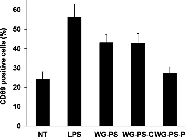
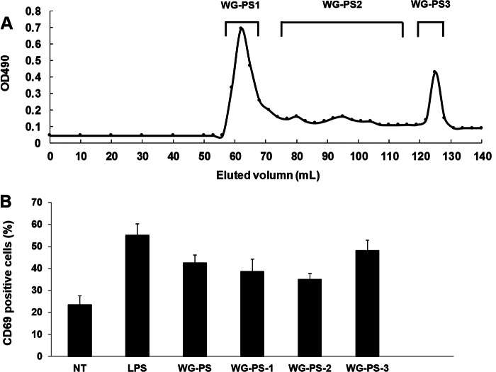
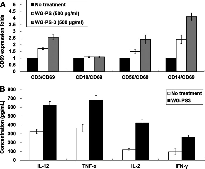
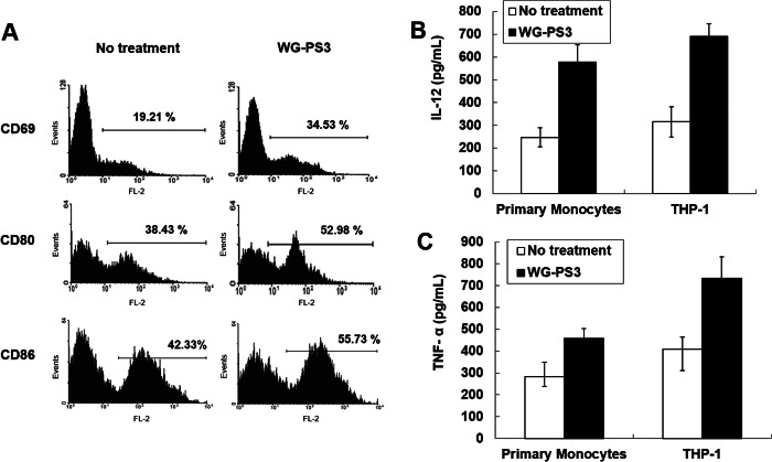
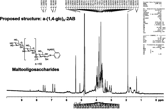
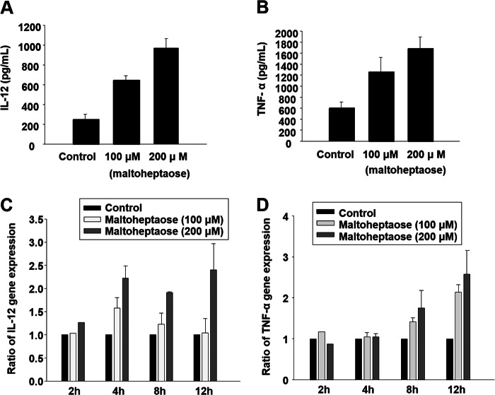
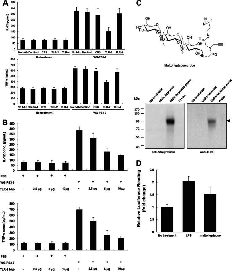
Wheatgrass is one of the most widely used health foods, but its functional components and mechanisms remain unexplored. Herein, wheatgrass-derived oligosaccharides (WG-PS3) were isolated and found to induce CD69 and Th1 cytokine expression in human peripheral blood mononuclear cells. In particular, WG-PS3 directly activated the purified monocytes by inducing the expression of CD69, CD80, CD86, IL-12, and TNF-α but affected NK and T cells only in the presence of monocytes. After further purification and structural analysis, maltoheptaose was identified from WG-PS3 as an immunomodulator. Maltoheptaose activated monocytes via Toll-like receptor 2 (TLR-2) signaling, as discovered by pretreatment of blocking antibodies against Toll-like receptors (TLRs) and also determined by click chemistry. This study is the first to reveal the immunostimulatory component of wheatgrass with well defined molecular structures and mechanisms.

Keywords: Carbohydrate Function; Immune Activation; Maltoheptaose; Monocytes; Natural Products; Oligosaccharide; Toll-like Receptors (TLR); Wheatgrass.


Figures

FIGURE 1.
FIGURE 1.
The wheatgrass extracts induce CD69 expression in hPBMCs. hPBMCs (1 × 10⁶ cells) were treated with LPS (positive control, 1 μg/ml) and wheatgrass extracts (500 μg/ml) separately for 24 h. The samples of wheatgrass extracts were as follows: WG-PS, WG-PS-C (the peptide-free WG-PS), and WG-PS-P (the carbohydrate-free WG-PS). The hPBMCs were stained with an anti-CD69-PE antibody and analyzed for the ratio of positive signals to isotype-negative controls. The percentage of hPBMCs with positive fluorescence signals were calculated and normalized to the treatment-free value. The results are expressed as the mean ± S.E. (error bars) from three independent experiments. NT, no treatment.
FIGURE 2.
FIGURE 2.
The fraction WG-PS3 induces CD69 expression in hPBMCs. A, wheatgrass extracts were separated by size exclusion chromatography. B, hPBMCs (1 × 10⁶) were treated with each fraction (500 μg/ml) separately for 24 h. hPBMCs were then stained with an anti-CD69-PE antibody and analyzed for the percentage of positive signals. The percentage of hPBMCs with positive fluorescence signals was calculated and normalized to the treatment-free value. The results are expressed as the mean ± S.E. (error bars) from three independent experiments. NT, no treatment.
FIGURE 3.
FIGURE 3.
The effect of WG-PS3 on purified hPBMC subsets and Th1 cytokine secretions. A, 4 × 10⁶ hPBMCs were treated with WG-PS and WG-PS3 (500 μg/ml) and treated with PBS as the no treatment control. The double staining was performed after 24 h and detected by flow cytometry. The percentages of CD14+/CD69+, CD3+/CD69+, and CD56+/CD69+ cells were measured and normalized to the no treatment value. B, hPBMCs (1 × 10⁶) were treated with WG-PS3 (100 μg/ml). The conditioned medium was harvested at 24 h and assayed for the concentrations of IL-12, TNF-α, IL-2, and IFN-γ by ELISA (100 μl of conditioned medium/assay). The results are expressed as the mean ± S.E. (error bars) from three independent experiments.
FIGURE 4.
FIGURE 4.
WG-PS3 activates primary CD14+ monocytes and THP-1 cells. A, the primary CD14 monocytes (1 × 10⁶) were purified from hPBMCs by immunomagnetic negative selection and incubated with WG-PS3 (100 μg/ml) for 24 h, followed by expression analysis of the monocyte activation markers CD69, CD80, and CD86 using flow cytometry. B and C, the conditioned medium was collected, and the concentrations of IL-12 (B) and TNF-α (C) were measured by IL-12- and TNF-α-specific ELISA. The results are expressed as the mean ± S.E. (error bars) from three independent experiments.
FIGURE 5.
FIGURE 5.
The NMR spectra of WG-PS3-9.
FIGURE 6.
FIGURE 6.
Maltoheptaose induces IL-12 and TNF-α protein secretion and gene expression in THP-1 cells. A and B, THP-1 cells were treated with maltoheptaose (100 and 200 μm) for 24 h. The conditioned medium was harvested, and the concentrations of IL-12 and TNF-α were assayed by ELISA (100 μl of conditioned medium/assay). C and D, quantitative real-time PCR analysis of IL-12 and TNF-α expression in THP-1 cells after treating with maltoheptaose (100 and 200 μm) for 2, 4, 8, and 12 h. The relative quantification was determined by comparing with the control sample. The experiments were repeated three times. The results are expressed as mean ± S.E. (error bars).
FIGURE 7.
FIGURE 7.
The identification of receptors targeted by maltoheptaose on THP-1 cells. A, THP-1 cells (5 × 10⁵ cells) were pretreated with monoclonal blocking antibodies (5 μg) specific to Dectin-1, CR3, TLR-2, and TLR-4 and then incubated with WG-PS3-9 at a concentration of 100 μg/ml. After 24 h, the conditioned medium was harvested, and the concentrations of IL-12 and TNF-α were assayed by ELISA. B, THP-1 cells (5 × 10⁵ cells) were pretreated with blocking antibodies against TLR-2 (2, 5, and 10 μg) and then incubated with WG-PS3-9 at a concentration of 100 μg/ml. The conditioned medium was harvested after 24 h and analyzed for the concentrations of IL-12 and TNF-α using ELISA. The results are expressed as the mean ± S.E. (error bars) from three independent experiments. C, THP-1 cells (1 × 10⁸ cells) were untreated and treated with maltoheptaose, maltoheptaose-probe, or probe alone at a concentration of 8.67 mm for 2 h. After the reaction, diazirine photo-cross-linking and Cu(I)-catalyzed click chemistry were performed for covalently biotin labeling. Avidin beads were then employed for the purification of biotin-containing proteins that were resolved by electrophoresis and immunoblotting using streptavidin- and TLR2-specific antibodies. D, THP-1 cells were transiently transfected with NF-κB luciferase reporter plasmids followed by stimulation with maltoheptaose and the positive control, LPS, for 24 h, and the luciferase activity was then measured. The results are expressed as the mean ± S.E. from three independent experiments.

Similar articles

Cited by

References

    1. Alitheen N. B., Oon C. L., Keong Y. S., Chuan T. K., Li H. K., Yong H. W. (2011) Cytotoxic effects of commercial wheatgrass and fiber towards human acute promyelocytic leukemia cells (HL60). Pak. J. Pharm. Sci. 24, 243–250 - PubMed
    1. Das A., Raychaudhuri U., Chakraborty R. (2012) Effect of freeze drying and oven drying on antioxidant properties of fresh wheatgrass. Int. J. Food Sci. Nutr. 63, 718–721 - PubMed
    1. Ben-Arye E., Goldin E., Wengrower D., Stamper A., Kohn R., Berry E. (2002) Wheat grass juice in the treatment of active distal ulcerative colitis. A randomized double-blind placebo-controlled trial. Scand. J. Gastroenterol. 37, 444–449 - PubMed
    1. Shermer M. (2008) Wheatgrass juice and folk medicine. Sci. Am. 299, 42 - PubMed
    1. Chang R. (2002) Bioactive polysaccharides from traditional Chinese medicine herbs as anticancer adjuvants. J. Altern. Complement. Med. 8, 559–565 - PubMed

Publication types

MeSH terms