. 2013 Jun 14;288(24):17481-94.
doi: 10.1074/jbc.M112.439422. Epub 2013 Apr 29.

Molecular mechanism of 17-allylamino-17-demethoxygeldanamycin (17-AAG)-induced AXL receptor tyrosine kinase degradation

Affiliations

Molecular mechanism of 17-allylamino-17-demethoxygeldanamycin (17-AAG)-induced AXL receptor tyrosine kinase degradation

Gnana Prakasam Krishnamoorthy et al. J Biol Chem. .

Abstract

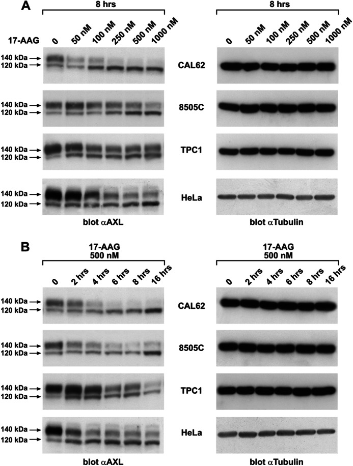
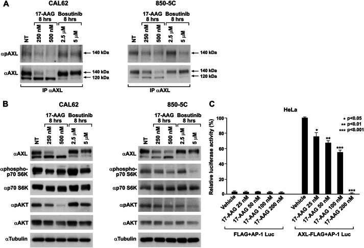
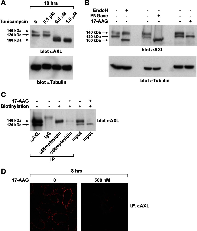
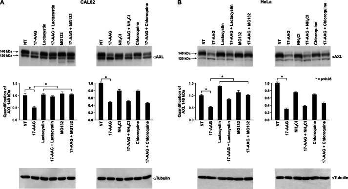
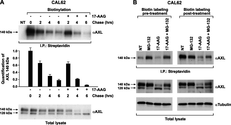
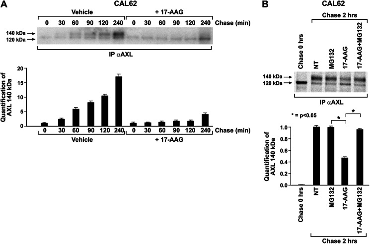
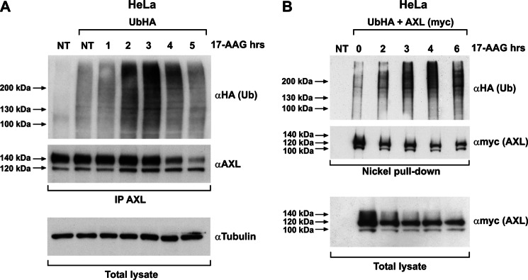
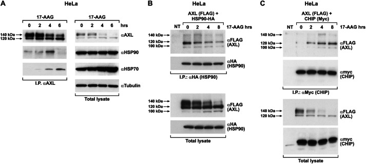
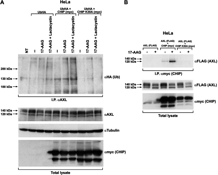
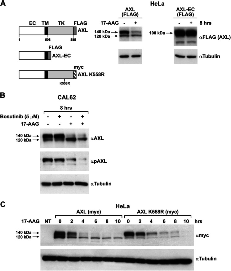
The receptor tyrosine kinase AXL is overexpressed in many cancer types including thyroid carcinomas and has well established roles in tumor formation and progression. Proper folding, maturation, and activity of several oncogenic receptor tyrosine kinases require HSP90 chaperoning. HSP90 inhibition by the antibiotic geldanamycin or its derivative 17-allylamino-17-demethoxygeldanamycin (17-AAG) causes destabilization of its client proteins. Here we show that AXL is a novel client protein of HSP90. 17-AAG induced a time- and dose-dependent down-regulation of endogenous or ectopically expressed AXL protein, thereby inhibiting AXL-mediated signaling and biological activity. 17-AAG-induced AXL down-regulation specifically affected fully glycosylated mature receptor present on cell membrane. By using biotin and [(35)S]methionine labeling, we showed that 17-AAG caused depletion of membrane-localized AXL by mediating its degradation in the intracellular compartment, thus restricting its exposure on the cell surface. 17-AAG induced AXL polyubiquitination and subsequent proteasomal degradation; under basal conditions, AXL co-immunoprecipitated with HSP90. Upon 17-AAG treatment, AXL associated with the co-chaperone HSP70 and the ubiquitin E3 ligase carboxyl terminus of HSC70-interacting protein (CHIP). Overexpression of CHIP, but not of the inactive mutant CHIP K30A, induced accumulation of AXL polyubiquitinated species upon 17-AAG treatment. The sensitivity of AXL to 17-AAG required its intracellular domain because an AXL intracellular domain-deleted mutant was insensitive to the compound. Active AXL and kinase-dead AXL were similarly sensitive to 17-AAG, implying that 17-AAG sensitivity does not require receptor phosphorylation. Overall our data elucidate the molecular basis of AXL down-regulation by HSP90 inhibitors and suggest that HSP90 inhibition in anticancer therapy can exert its effect through inhibition of multiple kinases including AXL.

Keywords: 17-AAG/Geldanamycin; AXL Receptor Tyrosine Kinase; CHIP E3 Ligase; E3 Ubiquitin Ligase; HSP90 Inhibition; Hsp90; Proteasome; Receptor Tyrosine Kinase; Ubiquitination.


Figures

FIGURE 1.
FIGURE 1.
17-AAG induces AXL degradation. A, protein lysates from the indicated thyroid carcinoma cell lines (CAL62, 8505C, and TPC1) and HeLa cells that were subjected to vehicle (0) or 17-AAG treatment with the indicated doses for 8 h were immunoblotted for AXL. Tubulin immunoblotting were used as a loading control. B, protein lysates from the indicated thyroid carcinoma cell lines and HeLa cells treated with 500 nm 17-AAG and harvested at the indicated time points were immunoblotted using anti-AXL and anti-tubulin antibodies.
FIGURE 2.
FIGURE 2.
17-AAG inhibits AXL-mediated signaling and downstream activity. A, CAL62 and 8505C cells treated with the indicated doses of 17-AAG and bosutinib were harvested and lysed, and equal amounts of proteins were immunoprecipitated (IP) with anti-AXL and immunoblotted using anti-phospho-AXL (pAXL). NT, nontreated. B, CAL62 and 8505C cells treated with 17-AAG and bosutinib at varying doses were subjected to Western blotting. AKT and p70 S6 kinase immunoblotting was done with the respective phospho- and total antibodies. Tubulin immunoblotting was used as a loading control. NT, nontreated. C, HeLa cells were transiently transfected with AXL-FLAG-expressing vector and the AP1-Luc vector. pRL-null (a plasmid expressing the enzyme Renilla luciferase from R. reniformis) was used as an internal control. Firefly and Renilla luciferase activities are expressed as the percentage of residual activity of 17-AAG-treated cells with respect to untreated cells. Average results of three independent assays ± S.D. are indicated. The analysis of variance Bonferroni multiple comparison test was used to demonstrate statistical significance. *, p < 0.05; **, p < 0.01; ***, p < 0.001.
FIGURE 3.
FIGURE 3.
17-AAG targets the fully glycosylated isoform of AXL. A, lysates of CAL62 cells treated 18 h with the indicated doses of tunicamycin were immunoblotted for AXL and normalized with anti-tubulin. B, 50 μg of protein lysates of CAL62 cells preboiled for 5 min at 99 °C was subjected to overnight Endo H (0.01 unit) and PNGase F (1 unit) digestion at 37 °C. The enzyme-digested lysates were subjected to Western blotting for AXL and tubulin. C, CAL62 cells treated or not with 17-AAG (500 nm) for 8 h were subjected to surface protein biotin labeling. Cells were then lysed, immunoprecipitated (IP) using streptavidin-agarose resin, and immunoblotted for AXL. D, CAL62 cells grown on a coverslip were subjected to vehicle (0) or 17-AAG (500 nm) treatment for 8 h. Cells were then fixed and stained for immunofluorescence (IF) using AXL antibody targeting the extracellular domain. Representative microscopy images of untreated and 17-AAG-treated cells are presented.
FIGURE 4.
FIGURE 4.
Proteasomal inhibitors restored 17-AAG-induced mature AXL depletion. Lysates of CAL62 (A) and HeLa (B) cells co-treated with 17-AAG (500 nm) and the proteasomal inhibitors MG132 (10 μm)/lactacystin (5 μm) or lysosomal inhibitors NH4Cl (20 mm)/chloroquine (100 μm) were subjected to Western blotting for AXL. Tubulin immunoblotting was used as a loading control. The 140-kDa AXL signals in the indicated treatments were quantified by densitometry, and the relative change compared with nontreated cells (NT) was plotted. The values are representative of three independent experiments. Error bars represent S.D. *, p < 0.05.
FIGURE 5.
FIGURE 5.
Effect of 17-AAG on plasma membrane-localized AXL. A, CAL62 cell surface proteins were biotinylated and chased in the presence of vehicle or 17-AAG (500 nm), and comparable amounts of lysates were immunoprecipitated (I.P.) with streptavidin-agarose and immunoblotted for AXL. AXL immunoblotting of total cell lysates was used to compare total AXL levels. The biotinylated AXL was analyzed by densitometry, and the relative change in the signal was plotted compared with no chase (0), which was considered equal to 1. The results are representative of three independent experiments. Error bars represent S.D. NT, non biotinylated. B, CAL62 cells biotinylated before and after treatments with 17-AAG (500 nm), MG132 (10 μm), and 17-AAG + MG132 for 5 h were subjected to streptavidin immunoprecipitation, and the recovered biotinylated proteins were subjected to Western blotting for AXL. AXL and tubulin immunoblotting was performed on total lysate. NT, non-treated cells.
FIGURE 6.
FIGURE 6.
HSP90 inhibition blocked AXL transport to the cell surface. A, CAL62 cells pulsed with [35S]methionine for 15 min and chased with nonradioactive medium with or without 17-AAG (500 nm) for the indicated time points. Cells were lysed and subjected to AXL immunoprecipitation (IP), and the dried gel was subjected to standard autoradiography. The 140-kDa signal was analyzed by densitometry and graphically represented. The results are representative of three independent experiments. B, CAL62 cells pulsed with [35S]methionine for 15 min were harvested unchased (0 h) or were chased with nonradioactive medium in the presence of MG132 (10 μm), 17-AAG (500 nm), and their combination for 2 h. Cells lysates were immunoprecipitated for AXL followed by SDS-PAGE, and the dried gel was subjected to autoradiography and phosphorimaging. The quantification of 140-kDa AXL species was analyzed by densitometry. The non-treated cell (NT) signal was considered equal to 1. The results are representative of three independent experiments. Error bars represent S.D. *, p < 0.05.
FIGURE 7.
FIGURE 7.
17-AAG induces AXL ubiquitination. A, HeLa cells transiently transfected with HA-tagged ubiquitin (UbHA) were subjected to vehicle (NT) or 500 nm 17-AAG treatment for the indicated time points. Cell lysates were immunoprecipitated (IP) using anti-AXL and immunoblotted using anti-HA and -AXL. Tubulin immunoblotting on total lysate is shown to normalize the input. NT, nontransfected/nontreated. B, HeLa cells co-transfected with UbHA and His-tagged AXL (subcloned in pcDNA4/TO A His/myc) were treated or not with 500 nm 17-AAG for the indicated times. Cells were harvested, and comparable amounts of cell suspension were subjected to nickel affinity protein purification under denaturing conditions following the protocol described under “Materials and Methods.” Purified proteins were subjected to Western blotting using anti-HA (Ub) and anti-myc (AXL). Comparable aliquot of cell suspension from the inputs were lysed using standard procedure and showed as total lysate, immunoblotted for myc (AXL). NT, nontransfected.
FIGURE 8.
FIGURE 8.
17-AAG modulates AXL interaction with HSP90 and HSP70. A, HeLa cells treated with 500 nm 17-AAG for the indicated times, and equal amounts of protein lysate were immunoprecipitated (I.P.) using AXL antibody followed by immunoblotting with anti-HSP90 and -HSP70. AXL, HSP90, HSP70, and tubulin levels were monitored using the respective antibody by Western blotting performed using total lysate. B, HeLa cells co-transfected with AXL-FLAG and HSP90-HA vectors were treated with vehicle (−) and 500 nm 17-AAG for the indicated times. Equal amounts of protein lysate were immunoprecipitated using anti-HA (HSP90) followed by immunoblotting with anti-FLAG (AXL). FLAG and HA immunoblotting was also performed on total lysates. NT, non-transfected cells. Arrows indicate the 100-, 120-, and 140-kDa isoforms of the receptor. C, HeLa cells co-transfected with AXL-FLAG and CHIP-myc were treated or not (−) with 500 nm 17-AAG for the indicated times. Comparable protein aliquots were immunoprecipitated using anti-myc (CHIP) followed by immunoblotting with anti-FLAG (AXL). FLAG and myc immunoblotting was performed on total lysate. NT, non-transfected cells.
FIGURE 9.
FIGURE 9.
CHIP E3 ligase is responsible for AXL ubiquitination upon 17-AAG treatment. A, HeLa cells co-transfected with UbHA and CHIP WT or CHIP K30A were treated for 3 h with 500 nm 17-AAG with and without lactacystin (10 μm). Cells were lysed, and equal amounts of proteins were immunoprecipitated (I.P.) using AXL antibody and immunoblotted with anti-HA. AXL, myc, and tubulin immunoblotting was performed to monitor their levels in the total lysate. NT, non-transfected cells. “–”, nontreated. B, HeLa cells co-transfected with AXL-FLAG and myc-tagged CHIP WT or the mutant CHIP K30A were treated with 500 nm 17-AAG and subjected to myc (CHIP) immunoprecipitation. CHIP immunocomplexes on Western blotting were probed using anti-FLAG (AXL). FLAG and myc immunoblotting in total cell lysates was used to check AXL and CHIP levels, respectively.
FIGURE 10.
FIGURE 10.
The kinase domain but not the kinase activity sensitizes AXL to 17-AAG. A, FLAG-tagged AXL WT and AXL-EC mutant and myc-tagged AXL kinase-dead mutant (AXL K558R) constructs are schematically represented. TK, tyrosine kinase domain; TM, transmembrane domain. HeLa cells transiently transfected with FLAG-tagged AXL WT and AXL-EC expression vectors were treated with 500 nm 17-AAG (+) or vehicle (−) for 8 h, and the lysates were immunoblotted with anti-FLAG and anti-tubulin. B, CAL62 cells were treated with vehicle (−) or with 5 μm bosutinib in the presence or absence of 17-AAG (500 nm). Cell lysates were immunoblotted with anti-AXL, anti-phospho-AXL (pAXL), or anti-tubulin antibodies. C, HeLa cells transfected with myc-tagged AXL WT and AXL kinase-dead mutant (AXL K558R) were treated with 500 nm 17-AAG for the indicated time points. The lysates were subjected to Western blotting for myc and tubulin. NT, non-transfected cells.

Similar articles

Cited by

References

    1. O'Bryan J. P., Frye R. A., Cogswell P. C., Neubauer A., Kitch B., Prokop C., Espinosa R., 3rd, Le Beau M. M., Earp H. S., Liu E. T. (1991) Axl, a transforming gene isolated from primary human myeloid leukemia cells, encodes a novel receptor tyrosine kinase. Mol. Cell. Biol. 11, 5016–5031 - PMC - PubMed
    1. Stitt T. N., Conn G., Gore M., Lai C., Bruno J., Radziejewski C., Mattsson K., Fisher J., Gies D. R., Jones P. F., Masiakowski P., Ryan T. E., Tobkes N. J., Chen D. H., DiStefano P. S., Long G. L., Basilico C., Goldfarb M. P., Lemke G., Glass D. J., Yancopoulos G. D. (1995) The anticoagulation factor protein S and its relative, Gas6, are ligands for the Tyro 3/Axl family of receptor tyrosine kinases. Cell 80, 661–670 - PubMed
    1. Holland S. J., Pan A., Franci C., Hu Y., Chang B., Li W., Duan M., Torneros A., Yu J., Heckrodt T. J., Zhang J., Ding P., Apatira A., Chua J., Brandt R., Pine P., Goff D., Singh R., Payan D. G., Hitoshi Y. (2010) R428, a selective small molecule inhibitor of Axl kinase, blocks tumor spread and prolongs survival in models of metastatic breast cancer. Cancer Res. 70, 1544–1554 - PubMed
    1. Zhang Y. X., Knyazev P. G., Cheburkin Y. V., Sharma K., Knyazev Y. P., Orfi L., Szabadkai I., Daub H., Kéri G., Ullrich A. (2008) AXL is a potential target for therapeutic intervention in breast cancer progression. Cancer Res. 68, 1905–1915 - PubMed
    1. Rankin E. B., Fuh K. C., Taylor T. E., Krieg A. J., Musser M., Yuan J., Wei K., Kuo C. J., Longacre T. A., Giaccia A. J. (2010) AXL is an essential factor and therapeutic target for metastatic ovarian cancer. Cancer Res. 70, 7570–7579 - PMC - PubMed

Publication types

MeSH terms

Substances