. 2013 Jun 7;288(23):16882-16894.
doi: 10.1074/jbc.M112.422972. Epub 2013 Apr 29.

Myocilin stimulates osteogenic differentiation of mesenchymal stem cells through mitogen-activated protein kinase signaling

Affiliations

Myocilin stimulates osteogenic differentiation of mesenchymal stem cells through mitogen-activated protein kinase signaling

Heung Sun Kwon et al. J Biol Chem. .

Abstract

Myocilin is a secreted glycoprotein that is expressed in ocular and non-ocular tissues. Mutations in the MYOCILIN gene may lead to juvenile- and adult-onset primary open-angle glaucoma. Here we report that myocilin is expressed in bone marrow-derived mesenchymal stem cells (MSCs) and plays a role in their differentiation into osteoblasts in vitro and in osteogenesis in vivo. Expression of myocilin was detected in MSCs derived from mouse, rat, and human bone marrow, with human MSCs exhibiting the highest level of myocilin expression. Expression of myocilin rose during the course of human MSC differentiation into osteoblasts but not into adipocytes, and treatment with exogenous myocilin further enhanced osteogenesis. MSCs derived from Myoc-null mice had a reduced ability to differentiate into the osteoblastic lineage, which was partially rescued by exogenous extracellular myocilin treatment. Myocilin also stimulated osteogenic differentiation of wild-type MSCs, which was associated with activation of the p38, Erk1/2, and JNK MAP kinase signaling pathways as well as up-regulated expression of the osteogenic transcription factors Runx2 and Dlx5. Finally, cortical bone thickness and trabecular volume, as well as the expression level of osteopontin, a known factor of bone remodeling and osteoblast differentiation, were reduced dramatically in the femurs of Myoc-null mice compared with wild-type mice. These data suggest that myocilin should be considered as a target for improving the bone regenerative potential of MSCs and may identify a new role for myocilin in bone formation and/or maintenance in vivo.

Keywords: Differentiation; Glycoprotein Secretion; MAP Kinases (MAPKs); Mesenchymal Stem Cells; Myocilin; Osteogenesis; Signaling.


Figures

FIGURE 1.
FIGURE 1.
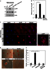
Myocilin expression in MSCs from different species. A, Western blot analysis of myocilin in lysates of MSCs from different species. Mouse skeletal muscle (mMuscle) was used as a positive control. Lysates were probed with polyclonal antibodies against mouse myocilin (1:2000). B, quantification of three independent Western blot experiments with the mean level of myocilin expression in mMSCs taken as one arbitrary unit. **, p < 0.01. C, relative levels of MYOC mRNA in hMSCs and human muscle as judged by qRT-PCR analysis. GAPDH mRNA was used for normalization. The mean level of MYOC expression in hMSCs was taken as one arbitrary unit. **, p < 0.01. D and E, intracellular distribution of myocilin in hMSCs. Cells were stained with antibodies against mouse myocilin (red) and antibodies against protein-disulfide isomerase (PDI) or golgin-97 (green). Nuclei were stained with DAPI (blue). The merged images demonstrate colocalization with endoplasmic reticulum (D) and Golgi markers (E). Typical immunofluorescence patterns are shown. Scale bar = 10 μm.
FIGURE 2.
FIGURE 2.
Changes in myocilin protein and MYOC mRNA in the course of MSC differentiation along the chondrogenic, adipogenic, and osteogenic lineages. A, Western blot analysis of myocilin in the lysates of undifferentiated (UD) hMSCs and hMSCs differentiating into chondrocytes (CH) after 2 weeks. Equal amounts of lysates were probed with antibodies against myocilin (1:2000) and HSC70 (1:5000). B, quantification of three independent Western blot experiments with the mean level of myocilin expression in undifferentiated hMSCs taken as one arbitrary unit. *, p < 0.05; **, p < 0.01. C, Western blot analysis of myocilin in the lysates of undifferentiated hMSCs and hMSCs differentiating into adipocytes (AD) or osteoblasts (OS) after 6 days. Equal amounts of lysates were probed with antibodies against myocilin (1:2000) and HSC70 (1:5000). D, quantification of three independent Western blot experiments with the mean level of myocilin expression in undifferentiated hMSCs taken as one arbitrary unit. **, p < 0.01. E, relative levels of MYOC mRNA in undifferentiated and differentiating hMSCs as judged by qRT-PCR analysis. hMSCs were differentiated for 4 days. GAPDH was used for normalization. The level of MYOC expression in undifferentiated hMSCs was taken as one arbitrary unit. **, p < 0.01.
FIGURE 3.
FIGURE 3.
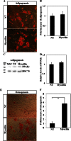
Effects of exogenous myocilin on hMSC differentiation along the adipogenic and osteogenic lineages. hMSCs were cultured for 6 days in adipogenic (A) or osteogenic (E) medium with or without addition of myocilin (3 μg/ml) and stained with Oil Red O or Alizarin Red S, respectively. NT, no treatment. B and F, quantification of the results shown in A and E. Four wells were counted per condition. The average number of cells counted per well was 350 and 470 in A and E, respectively. For statistical analysis, Student's t tests compared the average proportion of differentiated cells/well between the treatment and control groups, with n = 4 wells/group in each experiment. **, p < 0.01. Scale bar = 10 μm. C, Western blot analysis of the adipocyte marker PPARγ in the lysates of undifferentiated hMSCs (MSC) or hMSCs differentiating into adipocytes for 6 days in the absence (NT) or presence of myocilin (3 μg/ml). Equal amounts of lysates were probed with antibodies against PPARγ (1:2000) and HSC70 (1:5000). D, quantification of three independent Western blot experiments is shown, with the mean level of myocilin expression in undifferentiated hMSCs taken as one arbitrary unit.
FIGURE 4.
FIGURE 4.
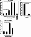
Dose dependence of myocilin in promoting osteogenesis in mMSCs from wild-type and Myoc-null mice. A, relative AP activity in mMSC culture lysates after cultivation of mMSCs in the presence of osteogenic induction medium plus increasing myocilin concentrations for 6 days. The level of AP activity in untreated mMSCs was taken as one arbitrary unit. **, p < 0.01. B, relative AP activity in mMSCs from wild-type and Myoc-null mice. AP activity was measured 6 days after initiation of osteoblastic differentiation. C, relative AP activity in Myoc-null mMSC culture lysates after cultivation of mMSCs in osteogenic differentiation medium and in the presence of increasing myocilin concentrations for 6 days. The level of AP activity in untreated mMSCs was taken as one arbitrary unit. Each panel depicts quantification of three independent experiments. **, p < 0.01.
FIGURE 5.
FIGURE 5.
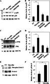
Myocilin-induced activation of the p38, JNK, and ERK signaling pathways. A, Western blot analysis of phosphorylated p38 (p-p38) levels in hMSCs differentiating into osteoblasts for 1 h in the absence (no treatment, NT) or presence of myocilin (3 μg/ml). SB203580 (10 μm), a specific inhibitor of p38, was added where shown. B, quantification of three independent Western blot experiments with the mean level of p-p38 normalized to total p38 in hMSCs differentiating without myocilin (NT) taken as one arbitrary unit. **, p < 0.01. C, Western blot analysis of phosphorylated JNK (p-JNK) levels in hMSCs differentiating into osteoblasts for 1 h in the absence (NT) or presence of myocilin (3 μg/ml). SP60125 (20 μm), a specific inhibitor of JNK, was added where shown. D, quantification of three independent Western blot experiments with the mean level of p-JNK normalized to total JNK in hMSCs differentiating without myocilin (NT) taken as one arbitrary unit. **, p < 0.01. E, Western blot analysis of phosphorylated Erk1/2 (p-Erk1/2) levels in hMSCs differentiating into osteoblasts for 1 h in the absence (NT) or presence of myocilin (3 μg/ml). F, quantification of three independent Western blot experiments with the mean level of p-Erk1/2 normalized to total Erk1/2 in hMSCs differentiating without myocilin taken as one arbitrary unit. **, p < 0.01. HSC70 was used for normalization in all experiments.
FIGURE 6.
FIGURE 6.
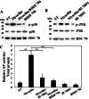
Inhibition of myocilin-induced activation of p38 and JNK as well as osteogenic differentiation by BIRB 796 and BI 78D3. Western blot analysis of phosphorylated p38 (p-p38) (A) and p-JNK (B) levels in hMSCs differentiating into osteoblasts for 1 h in the absence (no treatment, NT) or presence of myocilin (3 μg/ml). BIRB 796 (250 nm), a specific inhibitor of p38, and BI 78D3 (10 nm), a specific inhibitor of JNK, were added where shown. HSC70 was used for normalization in all experiments. C, relative AP activity in hMSCs measured 6 days after initiation of osteoblastic differentiation in the presence of myocilin (3 μg/ml), BIRB 796 (250 nm), and BI 78D3 (10 nm) is shown. Quantification of three independent experiments is shown, with the mean level of AP activity in hMSCs differentiating without myocilin (NT) taken as one arbitrary unit. **, p < 0.01.
FIGURE 7.
FIGURE 7.
Myocilin-induced activation of the p38 pathway. A, Western blot analysis of phosphorylated p38 (p-p38) levels in hMSCs differentiating into osteoblasts for 6 days in the absence (no treatment, NT) or presence of myocilin (3 μg/ml). Antiserum against myocilin was added to one sample. B, quantification of three independent Western blot experiments with the mean level of p-p38 in hMSC differentiating without myocilin taken as one arbitrary unit. **, p < 0.01. C, intracellular distribution of p-p38 (red) in hMSCs differentiating into osteoblasts in the absence (NT) and presence of myocilin (3 μg/ml) for 6 days. Nuclei were stained with DAPI (blue). Myocilin antiserum was added where shown. Scale bar = 5 μm. D and E, SB203580 inhibits myocilin-enhanced stimulation of hMSCs into osteoblasts. hMSCs were differentiated into osteoblasts in the absence (NT) or presence of myocilin (3 μg/ml). SB203580 (10 μm) was added where shown. Cells were stained with Alizarin Red S. Scale bar = 5 μm. Activity of AP was measured in cell lysates and normalized to total protein content. Shown is the quantification of three independent experiments, with the mean level of AP activity in hMSCs differentiating without myocilin taken as one arbitrary unit. **, p < 0.01.
FIGURE 8.
FIGURE 8.
Myocilin-induced activation of the Erk1/2 and JNK pathways. A, Western blot analysis of phosphorylated Erk1/2 (p-Erk1/2) levels in hMSCs differentiating into osteoblasts for 6 days in the absence (no treatment, NT) or presence of myocilin (3 μg/ml). B, quantification of three independent Western blot experiments is shown with the mean level of p-Erk1/2 in hMSC differentiating without myocilin taken as one arbitrary unit. **, p < 0.01. C, Western blot analysis of phosphorylated JNK (p-JNK) levels in undifferentiated hMSCs (Und) and hMSCs differentiating into osteoblasts for 6 days in the absence (NT) or presence of myocilin (3 μg/ml). D, quantification of three independent Western blot experiments with the mean level of p-JNK in hMSC differentiating without myocilin taken as one arbitrary unit. **, p < 0.01. E, SP600125 (20 μm) inhibits myocilin-enhanced stimulation of hMSCs into osteoblasts. Shown is the quantitation of three independent experiments, with the mean level of AP activity in hMSCs differentiating without myocilin for 6 days taken as one arbitrary unit. **, p < 0.01.
FIGURE 9.
FIGURE 9.
Myocilin-induced changes in the expression of Runx2 and Dlx5 associated with osteoblastic differentiation of MSCs. A, relative levels of indicated mRNAs in mMSCs differentiating into osteoblasts in the absence (no treatment, NT) or presence of myocilin (3 μg/ml) for 4 days as judged by qRT-PCR analysis. Antibodies against mouse myocilin were added where shown. GAPDH was used for normalization. The mean levels of indicated mRNAs in mMSCs from Myoc-null differentiating in the absence of myocilin were taken as one arbitrary unit. GAPDH mRNA was used for normalization. *, p < 0.05; **, p < 0.01. B, myocilin-induced changes in the level of Runx2 and Dlx5 protein during the course of osteoblastic differentiation of hMSCs (6 days) as judged by Western blot analysis. Equal amounts of lysates were probed with antibodies against Runx2 (1: 1000), Dlx5 (1:1000), and HSC70 (1:2000). These experiments were performed twice, and representative results are shown.
FIGURE 10.
FIGURE 10.
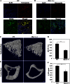
Analysis of bone structure in wild-type and Myoc-null mice. Immunostaining of frozen sections of the femur bone marrow from wild-type (A) and Myoc-null (B) mice with antibodies against osteopontin (1:500) and CD106 (1:500). Nuclei were stained with DAPI. Typical immunofluorescence patterns are shown. Scale bar = 10 μm. Representative three-dimensional reconstruction of the trabecular bone (C) and cortical bone (E) from femurs of 2-month-old wild-type and Myoc-null mouse littermates scanned using micro-CT. Micro-CT scans were performed as described under “Experimental Procedures.” Three pairs of mice were analyzed. Scale bar = 100 μm. Quantitative analyses of the representative three-dimensional reconstructions shown in C and E are presented in D and F, respectively. **, p < 0.01). TBV, trabecular bone volume; Ct.Th, midshaft cortical bone thickness.

Similar articles

Cited by

References

    1. Prockop D. J., Kota D. J., Bazhanov N., Reger R. L. (2010) Evolving paradigms for repair of tissues by adult stem/progenitor cells (MSCs). J. Cell Mol. Med. 14, 2190–2199 - PMC - PubMed
    1. English K., French A., Wood K. J. (2010) Mesenchymal stromal cells. Facilitators of successful transplantation? Cell Stem Cell, 7, 431–442 - PubMed
    1. Caplan A. I. (2007) Adult mesenchymal stem cells for tissue engineering versus regenerative medicine. J. Cell Physiol. 213, 341–347 - PubMed
    1. Pittenger M. F., Mackay A. M., Beck S. C., Jaiswal R. K., Douglas R., Mosca J. D., Moorman M. A., Simonetti D. W., Craig S., Marshak D. R. (1999) Multilineage potential of adult human mesenchymal stem cells. Science 284, 143–147 - PubMed
    1. Jiang Y., Jahagirdar B. N., Reinhardt R. L., Schwartz R. E., Keene C. D., Ortiz-Gonzalez X. R., Reyes M., Lenvik T., Lund T., Blackstad M., Du J., Aldrich S., Lisberg A., Low W. C., Largaespada D. A., Verfaillie C. M. (2002) Pluripotency of mesenchymal stem cells derived from adult marrow. Nature 418, 41–49 - PubMed

Publication types

MeSH terms