

doi: 10.3390/ijms14059475.
Oxidative stress mediates the disruption of airway epithelial tight junctions through a TRPM2-PLCγ1-PKCα signaling pathway
Affiliations
- PMID: 23629676
- PMCID: PMC3676794
- DOI: 10.3390/ijms14059475
Item in Clipboard
Oxidative stress mediates the disruption of airway epithelial tight junctions through a TRPM2-PLCγ1-PKCα signaling pathway
Int J Mol Sci.
.
Abstract
Oxidative stress has been implicated as an important contributing factor in the pathogenesis of several pulmonary inflammatory diseases. Previous studies have indicated a relationship between oxidative stress and the attenuation of epithelial tight junctions (TJs). In Human Bronchial Epithelial-16 cells (16HBE), we demonstrated the degradation of zonula occludens-1 (ZO-1), and claudin-2 exhibited a great dependence on the activation of the transient receptor potential melastatin (TRPM) 2 channel, phospholipase Cγ1 (PLCγ1) and the protein kinase Cα (PKCα) signaling cascade.
Figures
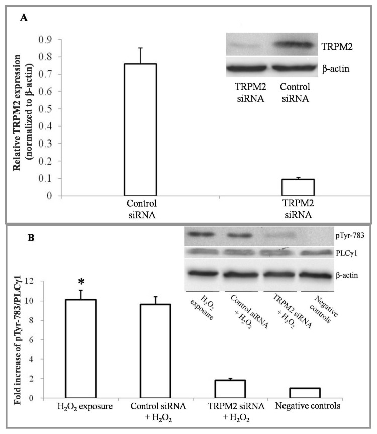
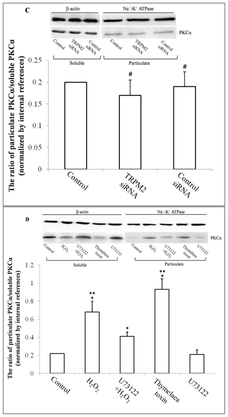
The activity of PLCγ1 and PKCα, estimated by Western blot analysis. (A) Compared to a control siRNA transfection, transient receptor potential melastatin (TRPM)-2 expression levels were reduced by >80% in the presence of a specific TRPM2 siRNA; (B) PLCγ1 and phosphorylated PLCγ1 at tyrosine 783 were detected by Western blot analysis. The protein levels were normalized with respect to β-actin. 16HBE cells treated with H2O2 free DMEM for 4 h were set as negative control. (n = 6 for each condition) * p < 0.05 for the TRPM2 siRNA transfection + H2O2 exposure group and negative controls; (C) PKCα was detected in both particulate and soluble extracts. To investigate whether TRPM2 depletion would influence the activity of PKCα, TRPM2 specific siRNA or control siRNA was transfected into 16HBE cells. (n = 6 for each condition), #p > 0.05 compared to the control; (D) PKCα was detected in both particulate and soluble extracts. 16HBE cells treated with H2O2-free DMEM for 4 h were set as negative control, (n = 6 for each condition), * p < 0.05, compared to the negative control, ** p < 0.05, compared to either the U73122 + H2O2 group or the negative control.
The activity of PLCγ1 and PKCα, estimated by Western blot analysis. (A) Compared to a control siRNA transfection, transient receptor potential melastatin (TRPM)-2 expression levels were reduced by >80% in the presence of a specific TRPM2 siRNA; (B) PLCγ1 and phosphorylated PLCγ1 at tyrosine 783 were detected by Western blot analysis. The protein levels were normalized with respect to β-actin. 16HBE cells treated with H2O2 free DMEM for 4 h were set as negative control. (n = 6 for each condition) * p < 0.05 for the TRPM2 siRNA transfection + H2O2 exposure group and negative controls; (C) PKCα was detected in both particulate and soluble extracts. To investigate whether TRPM2 depletion would influence the activity of PKCα, TRPM2 specific siRNA or control siRNA was transfected into 16HBE cells. (n = 6 for each condition), #p > 0.05 compared to the control; (D) PKCα was detected in both particulate and soluble extracts. 16HBE cells treated with H2O2-free DMEM for 4 h were set as negative control, (n = 6 for each condition), * p < 0.05, compared to the negative control, ** p < 0.05, compared to either the U73122 + H2O2 group or the negative control.
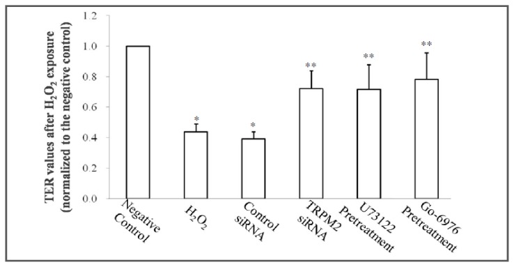
Effect of exogenous H2O2 individually or combined with TRPM2 siRNA, U73122 or Go-6976 on TER in 16HBE cells. TER values of each group after exposure to H2O2, were normalized to the average value of negative control after treatment with H2O2 free DMEM for 4 h, (n = 6 for each condition) * p < 0.05 compared to controls, ** p < 0.05 compared to the H2O2 group.
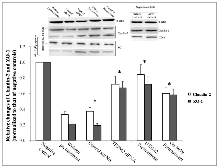
Expression levels of ZO-1 and claudin-2, the tight junction (TJ) protein family, were detected by western blot analysis. The results were normalized with respect to β-actin levels and adjusted to negative controls. 16HBE cells exposed to H2O2 free DMEM for 4 h were set as negative controls. (n = 6 for each condition), * p < 0.05 compared to the group without any pretreatments. #p > 0.05 compared to the group without any pretreatments.
Similar articles
-
Alcohol increases the permeability of airway epithelial tight junctions in Beas-2B and NHBE cells.Alcohol Clin Exp Res. 2012 Mar;36(3):432-42. doi: 10.1111/j.1530-0277.2011.01640.x. Epub 2011 Sep 26. Alcohol Clin Exp Res. 2012. PMID: 21950588 Free PMC article.
-
The degradation of airway tight junction protein under acidic conditions is probably mediated by transient receptor potential vanilloid 1 receptor.Biosci Rep. 2013 Oct 31;33(5):e00078. doi: 10.1042/BSR20130087. Biosci Rep. 2013. PMID: 24073800 Free PMC article.
-
Cigarette smoke-induced disruption of bronchial epithelial tight junctions is prevented by transforming growth factor-β.Am J Respir Cell Mol Biol. 2014 Jun;50(6):1040-52. doi: 10.1165/rcmb.2013-0090OC. Am J Respir Cell Mol Biol. 2014. PMID: 24358952
-
TRPM2 channel regulates endothelial barrier function.Adv Exp Med Biol. 2010;661:155-67. doi: 10.1007/978-1-60761-500-2_10. Adv Exp Med Biol. 2010. PMID: 20204729 Review.
-
The Epithelial Cell Leak Pathway.Int J Mol Sci. 2021 Jul 18;22(14):7677. doi: 10.3390/ijms22147677. Int J Mol Sci. 2021. PMID: 34299297 Free PMC article. Review.
Cited by
-
Dust particles-induced intracellular Ca2+ signaling and reactive oxygen species in lung fibroblast cell line MRC5.Korean J Physiol Pharmacol. 2017 May;21(3):327-334. doi: 10.4196/kjpp.2017.21.3.327. Epub 2017 Apr 21. Korean J Physiol Pharmacol. 2017. PMID: 28461775 Free PMC article.
-
Exposure to inhomogeneous static magnetic field beneficially affects allergic inflammation in a murine model.J R Soc Interface. 2014 Mar 19;11(95):20140097. doi: 10.1098/rsif.2014.0097. Print 2014 Jun 6. J R Soc Interface. 2014. PMID: 24647908 Free PMC article. Clinical Trial.
-
The role of stretch-activated ion channels in acute respiratory distress syndrome: finally a new target?Am J Physiol Lung Cell Mol Physiol. 2016 Sep 1;311(3):L639-52. doi: 10.1152/ajplung.00458.2015. Epub 2016 Aug 12. Am J Physiol Lung Cell Mol Physiol. 2016. PMID: 27521425 Free PMC article. Review.
-
Probiotic Properties and Safety Evaluation of Lactobacillus plantarum HY7718 with Superior Storage Stability Isolated from Fermented Squid.Microorganisms. 2023 Sep 8;11(9):2254. doi: 10.3390/microorganisms11092254. Microorganisms. 2023. PMID: 37764098 Free PMC article.
-
The Effect of Therapeutic Blockades of Dust Particles-Induced Ca²⁺ Signaling and Proinflammatory Cytokine IL-8 in Human Bronchial Epithelial Cells.Mediators Inflamm. 2015;2015:843024. doi: 10.1155/2015/843024. Epub 2015 Nov 10. Mediators Inflamm. 2015. PMID: 26640326 Free PMC article.
References
-
- Tomita K., Barnes P.J., Adcock I.M. The effect of oxidative stress on histone acetylation and IL-8 release. Biochem. Biophys. Res. Commun. 2003;301:572–577. - PubMed
-
- Van Itallie C.M., Anderson J.M. The molecular physiology of tight junction pores. Physiology. 2004;19:331–338. - PubMed
-
- Balda M.S., Matter K. Tight junctions and the regulation of gene expression. Biochim. Biophys. Acta. 2009;1788:761–767. - PubMed
-
- Wang F., Daugherty B., Keise L.L., Wei Z., Foley J.P., Savani R.C., Koval M. Heterogeneity of claudin expression by alveolar epithelial cells. Am. J. Respir. Cell Mol. Biol. 2003;29:62–70. - PubMed
Publication types
MeSH terms
Substances
LinkOut - more resources
Full Text Sources
Other Literature Sources
Molecular Biology Databases
Research Materials
