

Folate-based radiotracers for PET imaging--update and perspectives
- PMID: 23629756
- PMCID: PMC6269920
- DOI: 10.3390/molecules18055005
Folate-based radiotracers for PET imaging--update and perspectives
Abstract
The folate receptor (FR) is expressed in many tumor types, among those ovarian and lung cancer. Due to the high FR affinity of folic acid, it has been used for targeting of FR-positive tumors, allowing specific delivery of attached probes to the malignant tissue. Therefore, nuclear imaging of FR-positive cancer is of clinical interest for selecting patients who could benefit from innovative therapy concepts based on FR-targeting. Positron emission computed tomography (PET) has become an established technique in clinical routine because it provides an increased spatial resolution and higher sensitivity compared to single photon emission computed tomography (SPECT). Therefore, it is of critical importance to develop folate radiotracers suitable for PET imaging. This review article updates on the design, preparation and pre-clinical investigation of folate derivatives for radiolabeling with radioisotopes for PET. Among those the most relevant radionuclides so far are fluorine-18 (t(1/2): 110 min, E(av) β⁺: 250 keV) and gallium-68 (t(1/2): 68 min, E(av) β⁺: 830 keV). Recent results obtained with new PET isotopes such as terbium-152 (t(1/2): 17.5 h, Eβ⁺: 470 keV) or scandium-44 (t(1/2): 3.97 h, (Eav) β⁺: 632 keV) are also presented and discussed. Current endeavors for clinical implementation of PET agents open new perspectives for identification of FR-positive malignancies in patients.
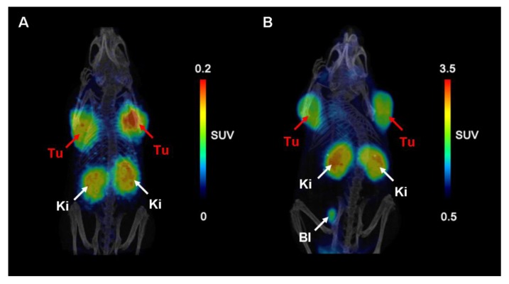
Figures
Similar articles
-
Synthesis and Preclinical Evaluation of Folate-NOTA-Al(18)F for PET Imaging of Folate-Receptor-Positive Tumors.Mol Pharm. 2016 May 2;13(5):1520-7. doi: 10.1021/acs.molpharmaceut.5b00989. Epub 2016 Apr 18. Mol Pharm. 2016. PMID: 27054811 Free PMC article.
-
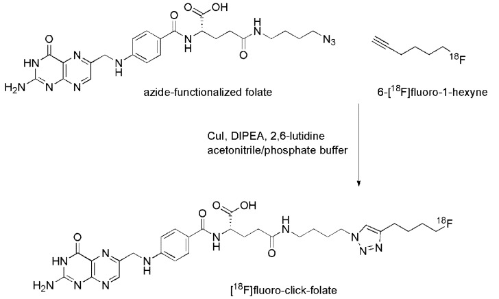
Fluorine-18 click radiosynthesis and preclinical evaluation of a new 18F-labeled folic acid derivative.Bioconjug Chem. 2008 Dec;19(12):2462-70. doi: 10.1021/bc800356r. Bioconjug Chem. 2008. PMID: 19053298
-
Reduced 18F-Folate Conjugates as a New Class of PET Tracers for Folate Receptor Imaging.Bioconjug Chem. 2018 Apr 18;29(4):1119-1130. doi: 10.1021/acs.bioconjchem.7b00775. Epub 2018 Feb 19. Bioconjug Chem. 2018. PMID: 29412638
-
Development of Folate Receptor-Targeted PET Radiopharmaceuticals for Tumor Imaging-A Bench-to-Bedside Journey.Cancers (Basel). 2020 Jun 9;12(6):1508. doi: 10.3390/cancers12061508. Cancers (Basel). 2020. PMID: 32527010 Free PMC article. Review.
-
(18) F-labeled folic acid derivatives for imaging of the folate receptor via positron emission tomography.J Labelled Comp Radiopharm. 2013 Jul-Aug;56(9-10):432-40. doi: 10.1002/jlcr.3104. J Labelled Comp Radiopharm. 2013. PMID: 24285516 Review.
Cited by
-
Contribution of Auger/conversion electrons to renal side effects after radionuclide therapy: preclinical comparison of (161)Tb-folate and (177)Lu-folate.EJNMMI Res. 2016 Dec;6(1):13. doi: 10.1186/s13550-016-0171-1. Epub 2016 Feb 9. EJNMMI Res. 2016. PMID: 26860295 Free PMC article.
-
Radiation dosimetry of 18F-AzaFol: A first in-human use of a folate receptor PET tracer.EJNMMI Res. 2020 Apr 8;10(1):32. doi: 10.1186/s13550-020-00624-2. EJNMMI Res. 2020. PMID: 32270313 Free PMC article.
-
Improving PET Quantification of Small Animal [68Ga]DOTA-Labeled PET/CT Studies by Using a CT-Based Positron Range Correction.Mol Imaging Biol. 2018 Aug;20(4):584-593. doi: 10.1007/s11307-018-1161-7. Mol Imaging Biol. 2018. PMID: 29352372
-
Development of a New Folate-Derived Ga-68-Based PET Imaging Agent.Mol Imaging Biol. 2017 Oct;19(5):754-761. doi: 10.1007/s11307-017-1049-y. Mol Imaging Biol. 2017. PMID: 28194631 Free PMC article.
-
Pharmacokinetic Properties of 68Ga-labelled Folic Acid Conjugates: Improvement Using HEHE Tag.Molecules. 2020 Jun 11;25(11):2712. doi: 10.3390/molecules25112712. Molecules. 2020. PMID: 32545327 Free PMC article.
References
-
- Gabriel M., Decristoforo C., Kendler D., Dobrozemsky G., Heute D., Uprimny C., Kovacs P., von Guggenberg E., Bale R., Virgolini I.J. 68Ga-DOTA-Tyr3-octreotide PET in neuroendocrine tumors: Comparison with somatostatin receptor scintigraphy and CT. J. Nucl. Med. 2007;48:508–518. doi: 10.2967/jnumed.106.035667. - DOI - PubMed
-
- Haug A.R., Auernhammer C.J., Wängler B., Schmidt G.P., Uebleis C., Goke B., Cumming P., Bartenstein P., Tiling R., Hacker M. 68Ga-DOTATATE PET/CT for the early prediction of response to somatostatin receptor-mediated radionuclide therapy in patients with well-differentiated neuroendocrine tumors. J. Nucl. Med. 2010;51:1349–1356. doi: 10.2967/jnumed.110.075002. - DOI - PubMed
Publication types
MeSH terms
Substances
LinkOut - more resources
Full Text Sources
Other Literature Sources
Medical
