. 2013 Jul;159(Pt 7):1254-1266.
doi: 10.1099/mic.0.066712-0. Epub 2013 Apr 29.

A novel approach to generate a recombinant toxoid vaccine against Clostridium difficile

Affiliations

A novel approach to generate a recombinant toxoid vaccine against Clostridium difficile

Robert G K Donald et al. Microbiology (Reading). 2013 Jul.

Abstract

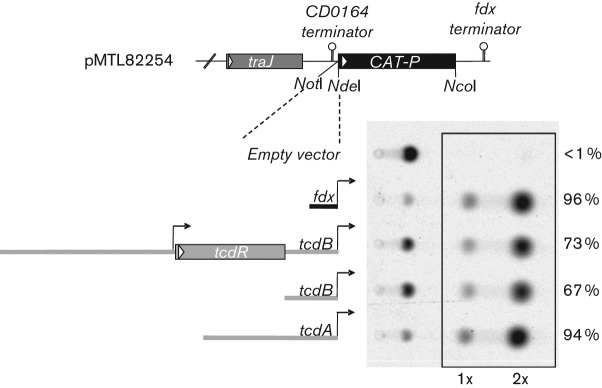
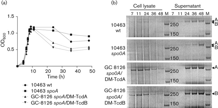
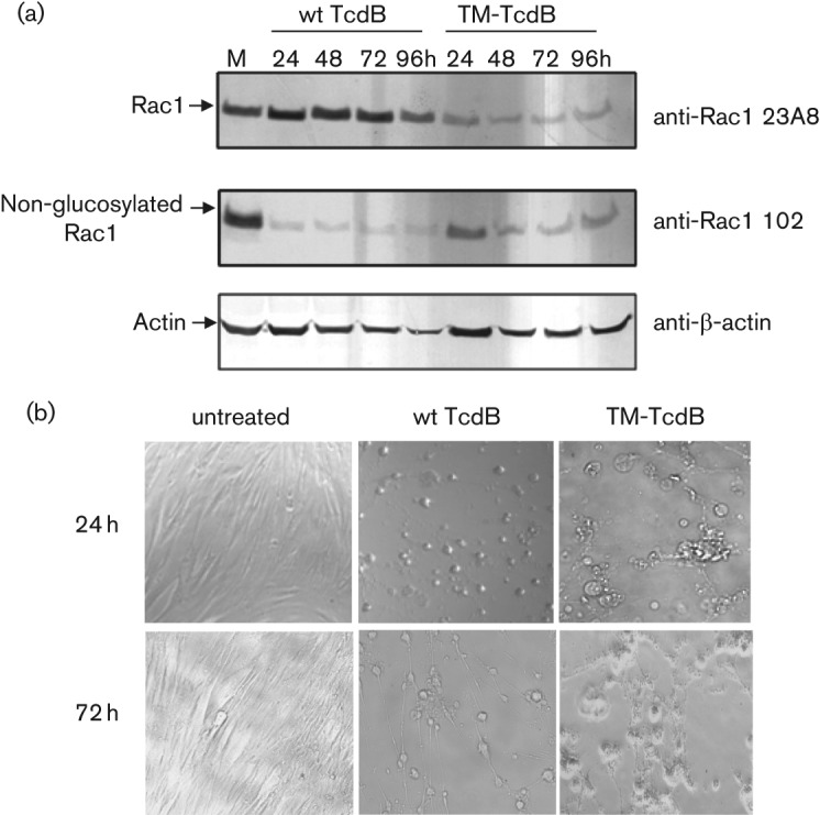
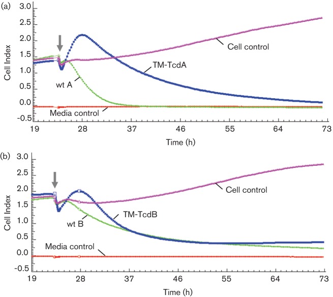
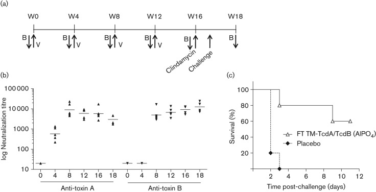
The Clostridium difficile toxins A and B are primarily responsible for symptoms of C. difficile associated disease and are prime targets for vaccine development. We describe a plasmid-based system for the production of genetically modified toxins in a non-sporulating strain of C. difficile that lacks the toxin genes tcdA and tcdB. TcdA and TcdB mutations targeting established glucosyltransferase cytotoxicity determinants were introduced into recombinant plasmids and episomally expressed toxin mutants purified from C. difficile transformants. TcdA and TcdB mutants lacking glucosyltransferase and autoproteolytic processing activities were ~10 000-fold less toxic to cultured human IMR-90 cells than corresponding recombinant or native toxins. However, both mutants retained residual cytotoxicity that could be prevented by preincubating the antigens with specific antibodies or by formalin treatment. Such non-toxic formalin-treated mutant antigens were immunogenic and protective in a hamster model of infection. The remaining toxicity of untreated TcdA and TcdB mutant antigens was associated with cellular swelling, a phenotype consistent with pore-induced membrane leakage. TcdB substitution mutations previously shown to block vesicular pore formation and toxin translocation substantially reduced residual toxicity. We discuss the implications of these results for the development of a C. difficile toxoid vaccine.


Figures

Fig. 1.
Fig. 1.
Evaluation of promoters for plasmid-based gene expression. C. difficile toxin and C. sporogenes fdx gene promoter fragments were subcloned in front of the CAT reporter in vector pMTL82254 (Heap et al., 2009) and plasmid constructs transferred by conjugation to C. difficile strain 630ΔErm. Transcriptional terminators, traJ conjugal transfer gene and flanking NotI and NdeI cloning sites are also illustrated. Crude total protein (5 µg) from lysates of stationary phase cultures were incubated with 14C-chloramphenicol and acetyl-CoA for 30 min at room temperature. CAT activity was determined by PhosphorImager analysis of radiolabelled components resolved by TLC. Mono- and bi-acetylated products are highlighted and the per cent conversion from un-acetylated substrate is indicated for each construct.
Fig. 2.
Fig. 2.
TcdA and TcdB expression in wt and recombinant toxin production strains. (a) Bacterial growth curves of strains expressing native toxins (VPI 10463, solid lines) and recombinant double mutant (DM) antigens (GC-8126, dotted lines). (b) TcdA and TcdB expression in cell pellets and in culture supernatants at 7, 11, 24, 36 and 48 h time points. Crude cell lysates and culture supernatants were separated by SDS-PAGE and high molecular mass toxins/toxoids visualized with Coomasie stain. Samples were concentrated 10-fold by membrane filtration before loading the gel. Arrowheads indicate location of full-length TcdA and TcdB proteins. M, molecular mass markers (250 and 150 kDa).
Fig. 3.
Fig. 3.
Structural features and GT activities of TM toxins. (a) Schematic highlighting mutations targeting GT catalytic and auto-proteolytic processing activities, modified from illustration in Jank et al. (2007a). Also indicated is the position of pore-inducing glutamate residues E970 and E976 within the TcdB hydrophobic region (HR). (b) InsP6-induced auto-proteolytic cleavage of purified wt TcdA/B and TM-TcdA/B toxins. Purified proteins (~1 µg) were incubated in the presence or absence of InsP6 at RT overnight. The cleavage products were separated by SDS-PAGE and stained with silver. Asterisks mark the molecular sizes of proteolytic cleavage products. (c) GT activity of native toxins and TM toxins. wt TcdA/B (1 ng) or TM-TcdA/B (100 µg) were incubated with RhoA GTPase in the presence of UDP-14C-glucose for 2 h at 30 °C. The activity of 100 µg TM-TcdA/B in the presence of 1 ng wt TcdA/B (wt+TM) was also assessed as control. The reaction mixture was resolved on SDS-PAGE and radioactive bands were visualized by PhosphorImager. Arrow indicates the 28 kDa 14C-labelled Rho GTPase protein band. (d) Concentration dependent toxin GT activity (at >0.1 ng ml−1) and lack of detectable activity (at 100 µg) for TM-antigens. After 2 h at 30 °C, reaction products were TCA precipitated and counted. Dotted line indicates the level of background 14C-glucose counts recovered from control reactions lacking TcdA or TcdB protein.
Fig. 4.
Fig. 4.
Neutralization of the cytotoxic activity of recombinant toxins with toxin-specific antibody or formalin pre-treatment. IMR-90 monolayers were incubated with serial dilutions of toxins and cell viability was determined after 3 days by measuring the release of intracellular ATP with a luciferase-based reagent. Error bars represent the standard deviations from results of duplicate serial dilutions. The relative cytotoxic activity of TM-TcdA (a) and TM-TcdB (b), with or without formalin or antibody pre-treatment, is illustrated. Neutralization of cytotoxic activity was achieved with a 1 : 10 dilution of toxin-specific rabbit polyclonal sera and by formalin pre-treatment.
Fig. 5.
Fig. 5.
The toxicity of TM-TcdB is not associated with glucosylation of intracellular Rac1 GTPase and is phenotypically distinct from wt TcdB-induced cytotoxicity. (a) Lysates from IMR-90 cells exposed for different times to wt TcdB (0.5 ng ml−1) or TM-TcdB (100 ng ml−1) and mock-treated cells (M, 24 h) were separated by SDS-PAGE. Protein bands were transferred to nitrocellulose and probed with anti-Rac1 monoclonal antibodies which recognize either all forms of Rac1 protein (mAb 23A8) or only the non-glucosylated form of Rac1 (mAb 102). Actin levels were monitored as internal control. (b) Cellular morphology of cells at 24 h and 72 h time points. TM-TcdB induces cellular swelling rather than the rounding phenotype that is characteristic of wt TcdB toxin.
Fig. 6.
Fig. 6.
Real-time xCelligence impedence analysis of toxin-induced cytotoxicity. IMR-90 cells growing on a microelectrode matrix were incubated with native and recombinant toxins and cellular impedence monitored every 15 min for 3 days. Media control traces represent culture medium without IMR-90 cells, while cell control traces are recordings of cells without toxin treatment. (a) Treatment with 0.1 µg wt TcdA ml−1 or 250 µg TM-TcdA ml−1. (b) Treatment with 0.1 ng wt TcdB ml−1 or 100 ng TMTcdB ml−1. The solid arrows indicate the time when toxins were added to the cell monolayer.
Fig. 7.
Fig. 7.
Immunogenicity and vaccine-induced protection in hamsters with formalin-treated TM-TcdA and TM-TcdB toxoids. (a) Groups of five Syrian golden hamsters were vaccinated (V) with 10 µg of each antigen in AlP04 or saline placebo via intramuscular injection at weeks 0, 4, 8 and 12. Animals were bled for serum samples at indicated time points (B). Clindamycin was given orally at week 16 and animals challenged five days later with 5×10³ c.f.u. of C. difficile strain 630. (b) Anti-toxin A and B neutralization titres were determined at the indicated weeks (x-axis) using a toxin neutralization assay. (c) Hamsters were monitored for CDAD severity using a clinical scoring system. Animals with total severity scores ≥15 were euthanized. The study terminated at day 11 post-challenge.

References

    1. Antunes A., Camiade E., Monot M., Courtois E., Barbut F., Sernova N. V., Rodionov D. A., Martin-Verstraete I., Dupuy B. (2012). Global transcriptional control by glucose and carbon regulator CcpA in Clostridium difficile. Nucleic Acids Res 40, 10701–10718. 10.1093/nar/gks864 - DOI - PMC - PubMed
    1. Barth H., Pfeifer G., Hofmann F., Maier E., Benz R., Aktories K. (2001). Low pH-induced formation of ion channels by Clostridium difficile toxin B in target cells. J Biol Chem 276, 10670–10676. 10.1074/jbc.M009445200 - DOI - PubMed
    1. Braun V., Hundsberger T., Leukel P., Sauerborn M., von Eichel-Streiber C. (1996). Definition of the single integration site of the pathogenicity locus in Clostridium difficile. Gene 181, 29–38. 10.1016/S0378-1119(96)00398-8 - DOI - PubMed
    1. Busch C., Hofmann F., Selzer J., Munro S., Jeckel D., Aktories K. (1998). A common motif of eukaryotic glycosyltransferases is essential for the enzyme activity of large clostridial cytotoxins. J Biol Chem 273, 19566–19572. 10.1074/jbc.273.31.19566 - DOI - PubMed
    1. Chumbler N. M., Farrow M. A., Lapierre L. A., Franklin J. L., Haslam D., Goldenring J. R., Lacy D. B. (2012). Clostridium difficile Toxin B causes epithelial cell necrosis through an autoprocessing-independent mechanism. PLoS Pathog 8, e1003072. 10.1371/journal.ppat.1003072 - DOI - PMC - PubMed

Publication types

MeSH terms

LinkOut - more resources