Review
. 2013 Apr 29;201(3):361-72.
doi: 10.1083/jcb.201302044.

Stress granules as crucibles of ALS pathogenesis

Affiliations
Review

Stress granules as crucibles of ALS pathogenesis

Yun R Li et al. J Cell Biol. .

Abstract

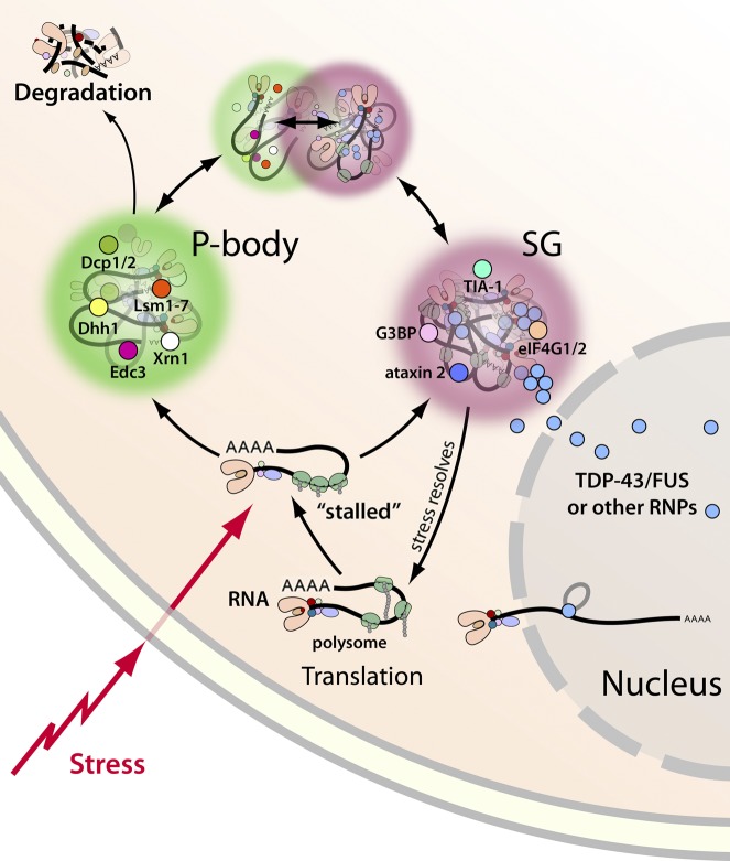
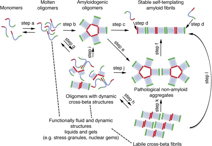
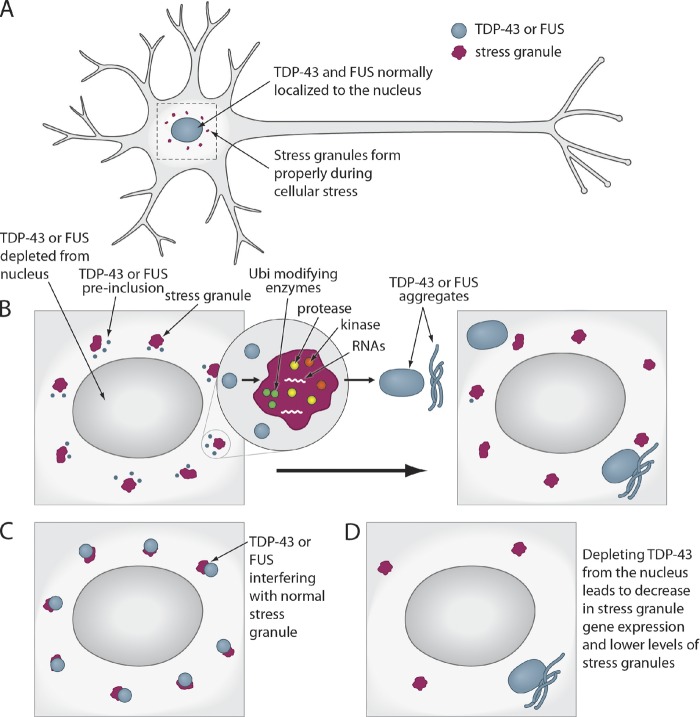
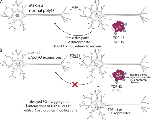
Amyotrophic lateral sclerosis (ALS) is a fatal human neurodegenerative disease affecting primarily motor neurons. Two RNA-binding proteins, TDP-43 and FUS, aggregate in the degenerating motor neurons of ALS patients, and mutations in the genes encoding these proteins cause some forms of ALS. TDP-43 and FUS and several related RNA-binding proteins harbor aggregation-promoting prion-like domains that allow them to rapidly self-associate. This property is critical for the formation and dynamics of cellular ribonucleoprotein granules, the crucibles of RNA metabolism and homeostasis. Recent work connecting TDP-43 and FUS to stress granules has suggested how this cellular pathway, which involves protein aggregation as part of its normal function, might be coopted during disease pathogenesis.


Figures

Figure 1.
Figure 1.
SGs and P-bodies are sites of RNA triage. Exposure to cellular stress can trigger a stress response that stalls translation initiation, resulting in the formation of SGs. SGs are dynamic cytoplasmic RNA–protein complexes that contain RNA-binding proteins, mRNAs, and translation initiation factors. When stress exposure dissipates, SGs disassemble and mRNA translation resumes. Nontranslating mRNAs can also be directed to P-bodies, distinct RNA-protein granules that are sites of stalled translation and mRNA degradation. SGs and P-bodies are differentially regulated and form independently, but they can and often do interact with each other. TDP-43, FUS, and other RNPs (e.g., TAF15, EWSR1, hnRNPA1, and hnRNPA2B1) reside predominantly in the nucleus, but stress exposure can trigger their recruitment to SGs. The implications for the recruitment of TDP-43, FUS, and others to SGs are explored in Fig. 3.
Figure 2.
Figure 2.
Prion and prion-like domains encode diverse conformational states and folding trajectories. Typically, prion and prion-like domains (depicted in blue, green, and red) populate a dynamic equilibrium comprised of soluble intrinsically unfolded monomers and molten oligomers (step a). These molten oligomers can subsequently evolve into multiple conformational states. In one trajectory, molten oligomers reorganize into more structured amyloidogenic oligomers (step b), which ultimately convert into a stable amyloid form (step c), which can self-template assembly (step d) and become infectious (i.e., a prion). Amyloidogenic oligomers can also cluster to form large pathological aggregates (step e), which might slowly convert to amyloid (step f). Alternatively, molten oligomers can partition into partially structured forms with dynamic cross-β structures that exhibit liquid-like properties (step g), and can rearrange further into labile cross-β fibrils with gel-like properties (step h). These liquid and gel-like collectives are likely critical structural components of various non–membrane-bound organelles including SGs and nuclear gems. Importantly, these transitions to liquid- and gel-like structures are readily reversible (steps a, g, and h). We suggest, however, that these structures are also prone to morph into amyloidogenic oligomers (step i), pathological nonamyloid aggregates (step j and k), and even stable self-templating amyloid (step l) connected with neurodegenerative disease.
Figure 3.
Figure 3.
How TDP-43 and FUS might interface with SGs during pathogenesis. (A) In normal neurons, TDP-43 and FUS are localized to the nucleus, and SGs form and dissipate normally. TDP-43 or FUS localization becomes abnormal during ALS pathogenesis and may interface with SGs in several different (and non-mutually exclusive) ways: (B) TDP-43 or FUS exit from the nucleus and begin to accumulate in the cytoplasm as preinclusions, where they interact and might colocalize with SGs. Here, TDP-43 and FUS might become modified by kinases, proteases, and ubiquitin-modifying enzymes, which are all present in SGs. These pathological modifications could accelerate TDP-43 and FUS aggregation or prevent return to the nucleus. Thus, SGs could serve as an obligate conduit for TDP-43 and FUS aggregation. Ubi, ubiquitin. (C) TDP-43 and FUS aggregation in the cytoplasm might interfere with SG function, perhaps by interfering with their ability to regulate RNAs targeted to these structures. (D) TDP-43 and FUS might be required in the nucleus for the regulation of SG genes, and therefore depletion from the nucleus could lead to a dysregulation of SG genes and decreased SG formation and function.
Figure 4.
Figure 4.
How ataxin 2 polyQ expansions might affect SGs in ALS. Ataxin 2 is a component of SGs and is required for their formation and function. Pathogenic polyQ expansions in ataxin 2 underlie spinocerebellar ataxia 2 and ALS. (A) SGs form upon cellular stress. The ataxin 2 polyQ length is normally 22 Q. When the stress dissipates, SGs dissolve. (B) In the presence of a pathogenic ataxin 2 polyQ expansion, the SGs might be more difficult to dissolve, perhaps owing to increased ataxin 2 stability. Persistent SGs would have a greater chance to interface with TDP-43 or FUS in the cytoplasm (TDP-43 and FUS normally shuttle in and out of nucleus). Thus, increased interactions of TDP-43 or FUS with SGs could lead to enhanced pathological modifications, resulting in TDP-43 or FUS aggregate formation.

Similar articles

Cited by

References

    1. Aguzzi A., Rajendran L. 2009. The transcellular spread of cytosolic amyloids, prions, and prionoids. Neuron. 64:783–790 10.1016/j.neuron.2009.12.016 - DOI - PubMed
    1. Alberti S., Halfmann R., King O., Kapila A., Lindquist S. 2009. A systematic survey identifies prions and illuminates sequence features of prionogenic proteins. Cell. 137:146–158 10.1016/j.cell.2009.02.044 - DOI - PMC - PubMed
    1. Anderson P., Kedersha N. 2008. Stress granules: the Tao of RNA triage. Trends Biochem. Sci. 33:141–150 10.1016/j.tibs.2007.12.003 - DOI - PubMed
    1. Arai T., Hasegawa M., Akiyama H., Ikeda K., Nonaka T., Mori H., Mann D., Tsuchiya K., Yoshida M., Hashizume Y., Oda T. 2006. TDP-43 is a component of ubiquitin-positive tau-negative inclusions in frontotemporal lobar degeneration and amyotrophic lateral sclerosis. Biochem. Biophys. Res. Commun. 351:602–611 10.1016/j.bbrc.2006.10.093 - DOI - PubMed
    1. Armakola M., Higgins M.J., Figley M.D., Barmada S.J., Scarborough E.A., Diaz Z., Fang X., Shorter J., Krogan N.J., Finkbeiner S., et al. 2012. Inhibition of RNA lariat debranching enzyme suppresses TDP-43 toxicity in ALS disease models. Nat. Genet. 44:1302–1309 10.1038/ng.2434 - DOI - PMC - PubMed

Publication types