. 2013 May 14;110(20):8004-9.
doi: 10.1073/pnas.1220143110. Epub 2013 Apr 29.

Differential hERG ion channel activity of ultrasmall gold nanoparticles

Affiliations

Differential hERG ion channel activity of ultrasmall gold nanoparticles

Annika Leifert et al. Proc Natl Acad Sci U S A. .

Abstract

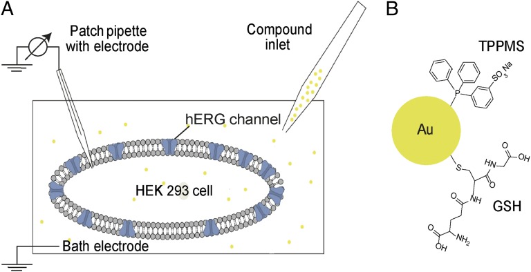
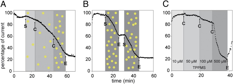
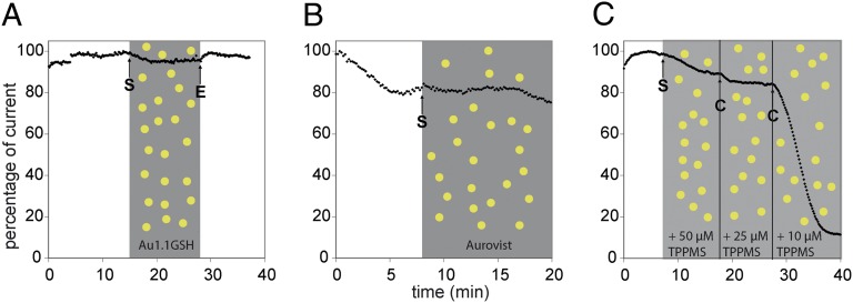
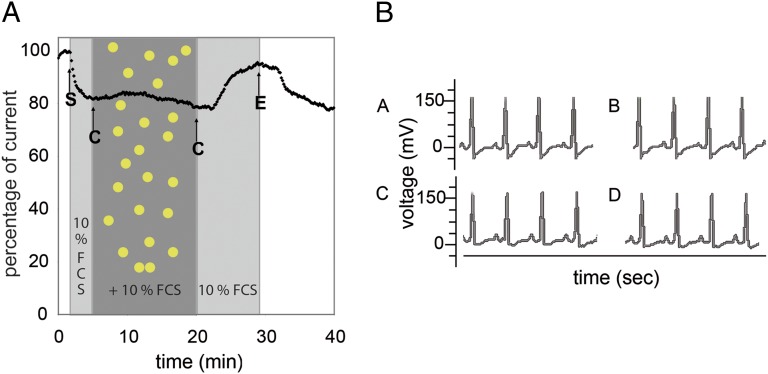
Understanding the mechanism of toxicity of nanomaterials remains a challenge with respect to both mechanisms involved and product regulation. Here we show toxicity of ultrasmall gold nanoparticles (AuNPs). Depending on the ligand chemistry, 1.4-nm-diameter AuNPs failed electrophysiology-based safety testing using human embryonic kidney cell line 293 cells expressing human ether-á-go-go-Related gene (hERG), a Food and Drug Administration-established drug safety test. In patch-clamp experiments, phosphine-stabilized AuNPs irreversibly blocked hERG channels, whereas thiol-stabilized AuNPs of similar size had no effect in vitro, and neither particle blocked the channel in vivo. We conclude that safety regulations may need to be reevaluated and adapted to reflect the fact that the binding modality of surface functional groups becomes a relevant parameter for the design of nanoscale bioactive compounds.

Keywords: complementarity; gold cluster; nanotoxicology; shape.


Conflict of interest statement

The authors declare no conflict of interest.

Figures

Scheme 1.
Scheme 1.
Schematic illustrations of the experimental setup and the molecular structure of tested species (not drawn to scale). (A) Scheme of patch-clamp setup in whole-cell configuration of a HEK 293 cell with hERG ion channels. Via the inlet, extracellular buffer (EC) or the respective test compound solution is perfused in the cell chamber. The connection between patch pipette and cell membrane is sealed to ensure a gigaohm seal, and the patch pipette is used to apply the voltage protocol as well as to record the response current. (B) AuNP surface with TPPMS ligand (with a AuNP core of 1.4 nm resulting in Au1.4MS) and GSH ligand (with a AuNP core of 1.1 nm resulting in Au1.1GSH), respectively.
Fig. 1.
Fig. 1.
Patch-clamp measurement of hERG tail current peak amplitudes in HEK 293 cells stably expressing the hERG ion channel. (A) Concentration-dependent inhibition of hERG current by Au1.4MS (3.1 µM, 6.5 µM, and 16.25 µM gold atom concentrations), sequentially applied to the same cell. The shaded areas indicate the intervals of different compound concentrations [Au] and [TPPMS], respectively. After a latency time of typically 2–3 min, an increase in the slope of current decay with increasing Au1.4MS concentration is observed. Arrows indicate start (S) and change (C) of perfusion with the differently concentrated AuNP solutions; E is end of sample perfusion (perfusion with EC). (B) Inhibition of hERG current by 65 µM Au1.4MS shows no recovery upon washout. Arrows indicate start (S) and end (E) of perfusion with AuNP solution. The blocking of the hERG channel by Au1.4MS is irreversible and additive. (C) Effect of TPPMS on the hERG tail current. Totals of 10 µM, 50 µM, 100 µM, and 500 µM TPPMS were subsequently perfused to the same cell. A total of 10 µM was applied from the start; arrows indicate change (C) and end (E) of sample perfusion. A total of 500 µM induces considerable channel inhibition, which is reversible, as opposed to blocking by Au1.4MS.
Fig. 2.
Fig. 2.
hERG tail current peak amplitudes in the presence of thiol-stabilized AuNPs or Au1.4MS with excess TPPMS. (A and B) Application of (A) 300 µM Au1.1GSH and (B) 300 µM AuroVist did not affect the hERG current. The shaded areas indicate the intervals of different compound concentrations of [Au] in Au1.1GSH (A), of [Au] in AuroVist (B), and of [TPPMS] (C), and the arrows indicate start (S) and end (E) of perfusion. (C) Preincubation of Au1.4MS with different concentrations of TPPMS abolished the hERG blocking potency of 20 µM Au1.4MS when TPPMS was present in excess. The cell was perfused with a mixture of 20 µM Au1.4MS + 50 µM TPPMS, 20 µM Au1.4MS + 25 µM TPPMS, and 20 µM Au1.4MS + 10 µM TPPMS. Arrows indicate start (S) and change (C) of sample perfusion.
Fig. 3.
Fig. 3.
Molecular simulation of nanoparticle docking to the intracellular hERG channel: (A) without TPPMS ligands [Au1.4MS(0)], (B) partially covered with 6 TPPMS [Au1.4MS(6)], and (C) with 12 TPPMS ligands [Au1.4MS(12)]. Shown are the molecular surface of the intracellular channel entrance (blue), the Au1.4 cluster in icosahedral form (yellow), and the ligands in element-specific color coding (C, green; O, red; S, yellow; P, orange; H, white). The dark red arrow in A indicates the entrance to the cavity on the intracellular side of the channel. This cavity can accommodate small-molecule ligands influencing hERG activity, but in our model not even the naked Au1.4 particle.
Fig. 4.
Fig. 4.
hERG tail current peak amplitudes in the presence of FCS and the representative mouse ECG waveforms recorded pre- and postinjection. The shaded areas indicate the intervals of FCS addition and Au1.4MS + FCS addition. (A) Application of 300 µM Au1.4MS in the presence of 10% FCS in the EC does not inhibit the hERG channel current. A slightly reduced tail current is observed with 10% FCS in EC (starting at arrow S) compared with EC without FCS. Other arrows indicate change (C) to Au1.4MS + 10% FCS and back to 10% FCS (after 20 min) and end (E) of sample perfusion. (B) ECG is taken before injection (A and C) and 6 d postinjection (B and D). The Mouse is injected with 0.9% NaCl (B) and 50 mg/kg Au1.4MS (D).

References

    1. Tiwari PM, Vig K, Dennis VA, Singh SR. Functionalized gold nanoparticles and their biomedical applications. Nanomaterials. 2011;1(1):31–63. - PMC - PubMed
    1. Daniel M-C, Astruc D. Gold nanoparticles: Assembly, supramolecular chemistry, quantum-size-related properties, and applications toward biology, catalysis, and nanotechnology. Chem Rev. 2004;104(1):293–346. - PubMed
    1. Giljohann DA, Mirkin CA. Drivers of biodiagnostic development. Nature. 2009;462(7272):461–464. - PMC - PubMed
    1. Kim B, et al. Tuning payload delivery in tumour cylindroids using gold nanoparticles. Nat Nanotechnol. 2010;5(6):465–472. - PMC - PubMed
    1. Popovtzer R, et al. Targeted gold nanoparticles enable molecular CT imaging of cancer. Nano Lett. 2008;8(12):4593–4596. - PMC - PubMed

Publication types

LinkOut - more resources