. 2013 May 14;110(20):8020-4.
doi: 10.1073/pnas.1221965110. Epub 2013 Apr 29.

Nanoscale analysis of pyritized microfossils reveals differential heterotrophic consumption in the ~1.9-Ga Gunflint chert

Affiliations

Nanoscale analysis of pyritized microfossils reveals differential heterotrophic consumption in the ~1.9-Ga Gunflint chert

David Wacey et al. Proc Natl Acad Sci U S A. .

Abstract

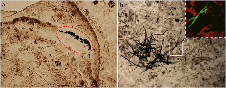
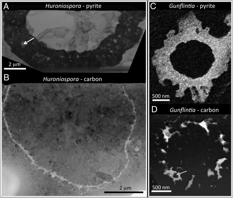
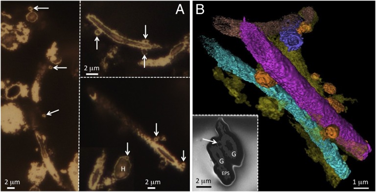
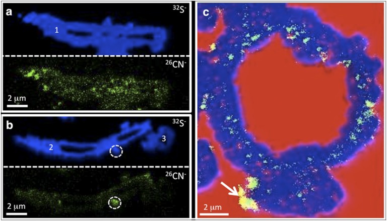
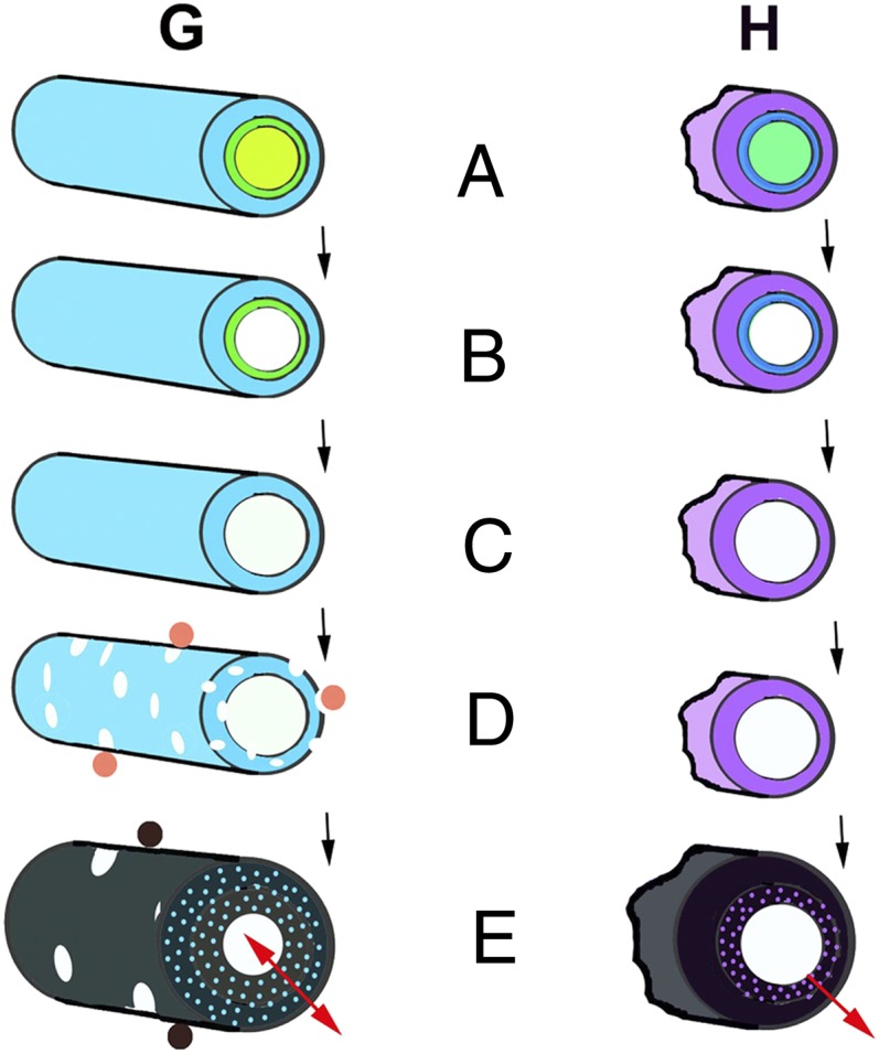
The 1.88-Ga Gunflint biota is one of the most famous Precambrian microfossil lagerstätten and provides a key record of the biosphere at a time of changing oceanic redox structure and chemistry. Here, we report on pyritized replicas of the iconic autotrophic Gunflintia-Huroniospora microfossil assemblage from the Schreiber Locality, Canada, that help capture a view through multiple trophic levels in a Paleoproterozoic ecosystem. Nanoscale analysis of pyritic Gunflintia (sheaths) and Huroniospora (cysts) reveals differing relic carbon and nitrogen distributions caused by contrasting spectra of decay and pyritization between taxa, reflecting in part their primary organic compositions. In situ sulfur isotope measurements from individual microfossils (δ(34)S(V-CDT) +6.7‰ to +21.5‰) show that pyritization was mediated by sulfate-reducing microbes within sediment pore waters whose sulfate ion concentrations rapidly became depleted, owing to occlusion of pore space by coeval silicification. Three-dimensional nanotomography reveals additional pyritized biomaterial, including hollow, cellular epibionts and extracellular polymeric substances, showing a preference for attachment to Gunflintia over Huroniospora and interpreted as components of a saprophytic heterotrophic, decomposing community. This work also extends the record of remarkable biological preservation in pyrite back to the Paleoproterozoic and provides criteria to assess the authenticity of even older pyritized microstructures that may represent some of the earliest evidence for life on our planet.

Keywords: biogeochemistry; paleontology; taphonomy.


Conflict of interest statement

The authors declare no conflict of interest.

Figures

Fig. 1.
Fig. 1.
Occurrence of pyritic microfossils at Schreiber Channel. (A) Stromatolitic chert with microfossil-rich laminae. Pyritic microfossils occur most commonly in millimeter-sized patches surrounded by clear chert (circled). These patches frequently pass laterally into areas rich in organic material and carbonaceous microfossils. (B) Laser Raman map (Inset) showing filamentous sheaths of Gunflintia that are part carbon (red) and part pyrite (green).
Fig. 2.
Fig. 2.
Changes in microfossil morphology and ultrastructure during pyritization. (A) Pyritized Huroniospora (bright-field TEM image) demonstrating thick (up to ∼2 μm) pyrite walls (dark gray) enclosing numerous nanograins of silica (pale gray; arrow). (B) Carbonaceous Huroniospora (bright-field TEM image) with thinner walls (mostly ∼200 nm) comprising a ring of carbon (white/pale gray) with a sawtooth texture caused by impinging silica nanograins. (C) Pyritized Gunflintia sheath (energy-filtered TEM image showing iron distribution) demonstrating thick pyrite walls (∼500 nm) and pyrite overgrowths. (D) Carbonaceous Gunflintia sheath (energy-filtered TEM image showing carbon distribution) showing poorer quality of preservation than Huroniospora, with walls comprising discontinuous rings of carbon. Nanograins of silica once again impinge upon and may be included within these walls (arrow). B and D were modified from (24), Copyright (2012) with permission from Elsevier.
Fig. 3.
Fig. 3.
Morphological evidence for saprophytic heterotrophs and EPSs. (A) Reflected light images of ellipsoidal–spheroidal epibionts up to ∼1 μm in diameter (arrows) attached to or embedded within Gunflintia and, more rarely, Huroniospora (H). (B) Three-dimensional reconstruction and visualization of the pyritic Gunflint biota (reconstructed from ∼80 individual FIB-SEM images spaced 75 nm apart). Micrometer-sized pyritic spheres/ellipses (orange) are attached to or embedded within pyritized Gunflintia sheaths and are interpreted here as prokaryotic, saprophytic heterotrophs. Other pyritic material (yellow) is attached or occurs close to Gunflintia sheaths; this has neither a crystalline nor cellular morphology and is interpreted here as pyritized EPSs. (Inset) Single FIB-SEM slice indicating the hollow nature of the pyritic Gunflintia sheaths (G) and saprophytic heterotroph (arrow), as well as inferred pyritized EPSs.
Fig. 4.
Fig. 4.
Chemical biosignals preserved within pyritic microfossils. (A and B) NanoSIMS ion images of sulfur (32S) and nitrogen (26CN) from three pyritic Gunflintia microfossils (–3). The pyrite frequently contains a chemical biosignal in the form of nitrogen enrichments. These are spatially variable both within an individual microfossil (1) and between microfossils (–3). Subcircular hotspots of nitrogen (dashed circle in B) frequently correlate with microspheroids observed in reflected light images, interpreted here as saprophytic heterotrophs. (C) Three-color overlay of NanoSIMS ion images from a pyritic Huroniospora microfossil: blue, pyrite; red, oxygen; and green, nitrogen. Note the discontinuous ring of nitrogen (green/yellow) within the pyritized microfossil, which is interpreted to represent a chemical ghost of the original organic microfossil wall. Hotspots of nitrogen exterior to the original wall (arrow) may represent mobilized organics from the original Huroniospora wall, the remains of saprophytic heterotrophs, or EPSs. Pores within the pyritic wall have been filled by nanograins of silica (red).
Fig. 5.
Fig. 5.
Typical trajectories of differential fossil decomposition and pyritization within the two dominant elements of the Gunflint microbiota seen at the Schreiber locality: prokaryotic sheaths of Gunflintia sp. (G) and prokaryotic cysts of Huroniospora sp. (H). Early diagenetic silicification has arrested microfossil decomposition at various stages (AE). (A) Sheaths and cysts still contain cell membranes with cytoplasmic contents (light green). (B) Cell cytoplasm plasmolyzed or decomposed, whereas the cell membrane remains relatively intact (green and blue inner rings). (C) The cell membrane decomposed, leaving only the sheath (G) and the cyst (H). (D) Aerobic heterotrophs (orange) break up the more labile sheath (G) but not the more refractory cyst (H). (E) Microbial sulfate reduction by anaerobic heterotrophic prokaryotes (brown) brings about pyritic replacement (gray-black) of both sheath (G) and cyst (H) material, involving dilation of carbonaceous and nitrogenous matter in the more labile sheath (G) or by marginal addition in the more refractory cyst (H).

References

    1. Gehling JG. Microbial mats in terminal Proterozoic siliciclastics: Ediacaran deathmasks. Palaios. 1999;14(1):40–57.
    1. Laflamme M, Schiffbauer JD, Narbonne GM, Briggs DEG. Microbial biofilms and the preservation of the Ediacara biota. Lethaia. 2011;44(2):203–213.
    1. Allison PA, Briggs DEG. 1991. Tissues. Taphonomy: Releasing the Data Locked in the Fossil Record, eds Allison PA, Briggs DEG (Plenum, New York), pp 25–69.
    1. Briggs DEG, Bottrell SH, Raiswell R. Pyritisation of soft-bodied fossils: Beecher’s Trilobite Bed, Upper Ordovician, New York State. Geology. 1991;19(12):1221–1224.
    1. Briggs DEG, Raiswell R, Bottrell SH, Hatfield D, Bartels C. Controls on the pyritization of exceptionally preserved fossils: An analysis of the Lower Devonian Hunsruck Slate of Germany. Am J Sci. 1996;296(6):633–663.

Publication types

LinkOut - more resources