. 2013 May 14;110(20):8146-51.
doi: 10.1073/pnas.1303126110. Epub 2013 Apr 29.

Mechanism of somatic hypermutation at the WA motif by human DNA polymerase η

Affiliations

Mechanism of somatic hypermutation at the WA motif by human DNA polymerase η

Ye Zhao et al. Proc Natl Acad Sci U S A. .

Abstract

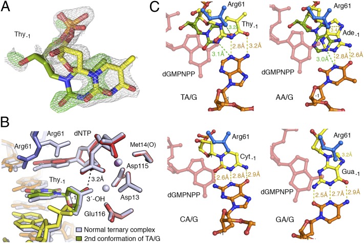
Somatic hypermutation is programmed base substitutions in the variable regions of Ig genes for high-affinity antibody generation. Two motifs, RGYW and WA (R, purine; Y, pyrimidine; W, A or T), have been found to be somatic hypermutation hotspots. Overwhelming evidence suggests that DNA polymerase η (Pol η) is responsible for converting the WA motif to WG by misincorporating dGTP opposite the templating T. To elucidate the molecular mechanism, crystal structures and kinetics of human Pol η substituting dGTP for dATP in four sequence contexts, TA, AA, GA, and CA, have been determined and compared. The T:dGTP wobble base pair is stabilized by Gln-38 and Arg-61, two uniquely conserved residues among Pol η. Weak base paring of the W (T:A or A:T) at the primer end and their distinct interactions with Pol η lead to misincorporation of G in the WA motif. Between two WA motifs, our kinetic and structural data indicate that A-to-G mutation occurs more readily in the TA context than AA. Finally, Pol η can extend the T:G mispair efficiently to complete the mutagenesis.

Keywords: A-to-G transition; immunoglobulin; π–cation stacking.


Conflict of interest statement

The authors declare no conflict of interest.

Figures

Fig. 1.
Fig. 1.
SHM at the WA motif. (A) Model of SHM targeting the WA motif through short-patch DNA synthesis. The initial incision targeting the variable region of Ig genes depends on AID and UNG. Recruitment of Pol η is enhanced by MutSα, which recognized the U:G mismatch. The template strand is in orange, and the primer strand (subject to mutation) is shown in yellow. The WA motif is highlighted with A in red. (B) Structural determination of the four stages of dGTP misincorporation in this study. The W of the WA motif is highlighted in yellow and the A subject to replacement by G in red. The protein domains of Pol η in contact with DNA and dNTP are outlined and labeled. The two Mg2+ ions in the active site are shown as purple spheres.
Fig. 2.
Fig. 2.
Structures of dGTP misincorporation by Pol η. (A) Structure superposition of Pol η complexed with DNA and nonhydrolyzable dGMPNPP opposite dT in all four sequence contexts. The protein is shown in ribbon diagrams with palm domain in pink, thumb in green, finger in blue, and little finger (LF) in magenta. DNA is shown as stick-and-ladder in yellow (primer) and orange (template). Mg2+ ions (purple spheres) and incoming dGMPNPP (red sticks) are also shown. (B) Correct (T:dATP) and incorrect (T:dGMPNPP) nascent base pair in complex with Pol η. The conserved residues Gln-38 and Arg-61 are shown in blue sticks. (C) Superposition of the Pol η active site in dGTP misincorporation (colored) and normal ternary complexes [gray; PDB ID code 3MR2]. Alterations of primer end and Lys-224 are indicated by red arrows. The water molecule that participates in Mg2+ coordination is shown as a red sphere. A gray double arrow indicates the cation–π stacking. (D) Superposition of the nascent (0) and (−1) base pairs in normal productive (gray) and mispaired nonproductive ternary complexes (colored). The movement of each nucleotide is indicated by red arrows.
Fig. 3.
Fig. 3.
The alternative DNA conformations observed in TA/G and AA/G that is compatible with dGTP incorporation. (A) The primer 3′ nucleotide (Thy) in the TA/G structure. The major (yellow) and the minor conformation (green) of the last nucleotide is superimposed with the 2FoFc (silver; contoured at 1.0σ) and FoFc omit map (green; contoured at 2.5σ), which were calculated without the minor conformation species. (B) The minor conformation of TA/G is compatible with the phosphoryltransfer reaction. Superposition of the minor conformation of the TA/G structure (colored) with the normal ternary complex (gray; PDB ID code 3MR2). (C) Interactions between Arg-61 (blue) and the −1 base pair (yellow, green primer, and orange template) in TA/G, AA/G, CA/G, and GA/G complexes. The incoming dGMPNPP is shown as semitransparent pink sticks. Hydrogen bonds and van der Waals contacts are indicated by color-coded dashes and distances.
Fig. 4.
Fig. 4.
Unique interactions found in TA/G and AA/G complexes. (A) Stereoview of the superposition of the four Pol η misincorporation and the normal ternary complexes. Each complex is color-coded as indicated. The upward shift of the DNA duplex in GA/G, CA/G, and AA/G complexes is obvious after the entire protein is superimposed. (B) Distinct interactions between the LF domain and the DNA found in TA/G and GA/G. The van der Waals contacts are indicated by lines formed by open circles. (C) Side-by-side views of TA/G and GA/G with Pol η shown as gray molecular surface and Ser-62 side chain in blue carbon and red oxygen. The shift of the DNA substrate in GA/G is accommodated by the rotamer change of Ser-62.
Fig. 5.
Fig. 5.
Translocation and extension of T:G mispair. (A) Structures of postinsertion Pol η–DNA binary complexes. The wobble nascent base pair of the misincorporation is shown side by side with the normal incorporation looking down the DNA helical axis. The template base is shown in orange and the newly incorporated base in yellow (correct) or red (incorrect). Arg-61 interacts with the mispaired guanine base. (B) Structures of posttranslocation binary complexes. In the T:G mismatch complex (Left), the DNA exhibits two equal populations of translocated (multicolored) and untranslocated (gray) conformations. The FoFc omit map, which was calculated with 100% translocated population and contoured at 2.5σ in green, superimposes well with the untranslocated conformation. (C) Structure of the T:G mismatch extension complex. The 2FoFc map (gray; contoured at 1.0σ) corresponding to the primer end and incoming nucleotide is superimposed with the refined structure. (D) Superposition of the T:G mismatch extension (colored) and normal ternary complex structure (gray; PDB ID code 3MR2). The 3′–OH at the primer end and the α-phosphate of dAMPNPP are aligned in the mismatch extension complex, albeit slightly shifted relative to the active site of Pol η.

References

    1. Rajewsky K, Förster I, Cumano A. Evolutionary and somatic selection of the antibody repertoire in the mouse. Science. 1987;238(4830):1088–1094. - PubMed
    1. Kim S, Davis M, Sinn E, Patten P, Hood L. Antibody diversity: Somatic hypermutation of rearranged VH genes. Cell. 1981;27(3 Pt 2):573–581. - PubMed
    1. Di Noia JM, Neuberger MS. Molecular mechanisms of antibody somatic hypermutation. Annu Rev Biochem. 2007;76:1–22. - PubMed
    1. Sohail A, Klapacz J, Samaranayake M, Ullah A, Bhagwat AS. Human activation-induced cytidine deaminase causes transcription-dependent, strand-biased C to U deaminations. Nucleic Acids Res. 2003;31(12):2990–2994. - PMC - PubMed
    1. Muramatsu M, Nagaoka H, Shinkura R, Begum NA, Honjo T. Discovery of activation-induced cytidine deaminase, the engraver of antibody memory. Adv Immunol. 2007;94:1–36. - PubMed

Publication types

LinkOut - more resources