Review
. 2013 Apr 24:7:76.
doi: 10.3389/fncir.2013.00076. eCollection 2013.

Imaging zebrafish neural circuitry from whole brain to synapse

Affiliations
Review

Imaging zebrafish neural circuitry from whole brain to synapse

Louis C Leung et al. Front Neural Circuits. .

Abstract

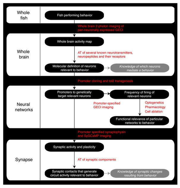
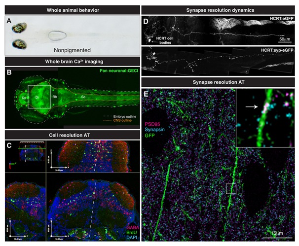
Recent advances in imaging tools are inspiring zebrafish researchers to tackle ever more ambitious questions in the neurosciences. Behaviorally fundamental conserved neural networks can now be potentially studied using zebrafish from a brain-wide scale to molecular resolution. In this perspective, we offer a roadmap by which a zebrafish researcher can navigate the course from collecting neural activities across the brain associated with a behavior, to unraveling molecular identities and testing the functional relevance of active neurons. In doing so, important insights will be gained as to how neural networks generate behaviors and assimilate changes in synaptic connectivity.

Keywords: array tomography; calcium imaging; clinical neuroscience; psychiatry; synapse imaging; whole brain imaging; zebrafish.


Figures

FIGURE 1
FIGURE 1
Comparison of brain sizes of common vertebrate neuroscience models. (A) Schematic diagram of the lateral view of adult human, mouse, and zebrafish brains to scale. (B, C) Schematic diagram of the linear organization of major brain regions from olfactory bulb to spinal cord (B) and the dorsal location of telencephalic structures such as the presumptive hippocampus and amygdala (C; adapted from Mueller et al., 2011). OB, olfactory bulb; Tel, Telencephalon; Di, Diencephalon; Mes, Mesencephalon; Rh, Rhombencephalon; sc, Spinal cord; Ctx, Neocortex; Hip, Hippocampus, pirCtx, Piriform cortex; BLA, Basolateral Amygdala and lot, lateral olfactory tract.
FIGURE 2
FIGURE 2
Roadmap of the steps a zebrafish neuroscience researcher can take to integrate the various levels of analysis within the same model organism. GECI, Genetically encoded calcium indicator; AT, Array Tomography; tol2, Tol2 transposase; SyGCaMP, Synaptophysin-specific GCaMP sensor.
FIGURE 3
FIGURE 3
From whole brain to single synapse. (A) Behavioral processes can be studied in non-pigmented (Nacre; mitfa mutant) species of zebrafish. (B) Two-photon volume image of 5 dpf zebrafish expressing GCaMP pan-neuronally, note the linear organization of the brain along the rostrocaudal axis from telencephalon (tel) to spinal cord (sc). (C) Various maximal projections (from area indicated in B by white box) of the 5 dpf zebrafish midbrain reconstructed after array tomography against GABA (magenta), BrdU (green) and DAPI (cyan) markers. White dashed line depicts midline. (D) Genetically specified circuit analysis. Synaptophysin-eGFP (syp-eGFP) allows imaging of structural plasticity of synapses (white arrowheads) in hypocretin (HCRT) neurons. (E) Single synapse resolution projection of array tomography sections stained for GFP (green), PSD95 (magenta) and Synapsin (cyan). Inset, magnification of a single shaft synapse (white arrow).

Similar articles

Cited by

References

    1. Ahrens M. B., Keller P. J. (2013). Whole-brain functional imaging at cellular resolution using light-sheet microscopy. Nat. Methods 10.1038/nmeth.2434 [Epub ahead of print]. - DOI - PubMed
    1. Ahrens M. B., Li J. M., Orger M. B., Robson D. N., Schier A. F., Engert F., et al. (2012). Brain-wide neuronal dynamics during motor adaptation in zebrafish. Nature 485 471–477. - PMC - PubMed
    1. Akanji A. O., George A. O., Olasode B. J., Osotimehin B. O. (1990). Fasting hypoglycaemia due to insulinoma in pregnancy. Postgrad. Med. J. 66 156. - PMC - PubMed
    1. Akerboom J., Carreras Calderon N., Tian L., Wabnig S., Prigge M., Tolo J., et al. (2013). Genetically encoded calcium indicators for multi-color neural activity imaging and combination with optogenetics. Front. Mol. Neurosci. 6:2 10.3389/fnmol.2013.00002 - DOI - PMC - PubMed
    1. Akerboom J., Chen T. W., Wardill T. J., Tian L., Marvin J. S., Mutlu S., et al. (2012). Optimization of a GCaMP calcium indicator for neural activity imaging. J. Neurosci. 32 13819–13840. - PMC - PubMed

Publication types

LinkOut - more resources