

White-matter changes correlate with cognitive functioning in Parkinson's disease
- PMID: 23630517
- PMCID: PMC3624087
- DOI: 10.3389/fneur.2013.00037
White-matter changes correlate with cognitive functioning in Parkinson's disease
Abstract
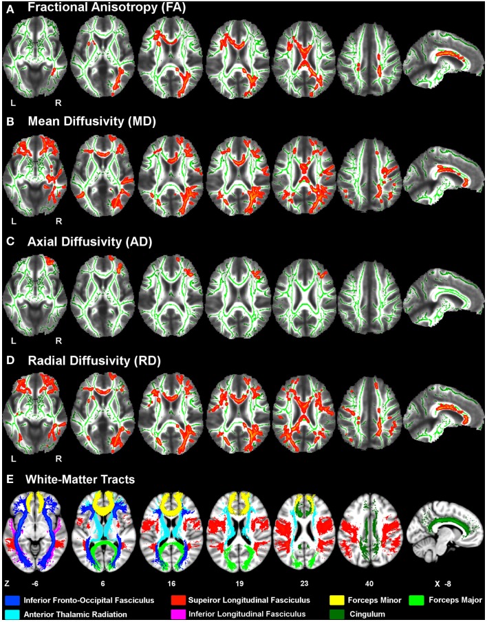
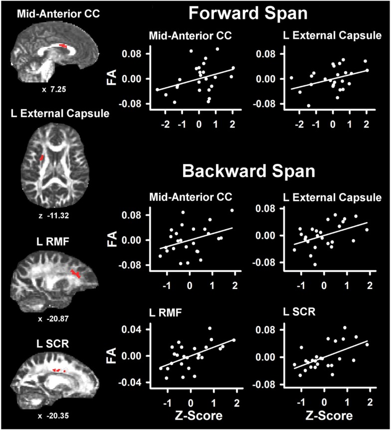
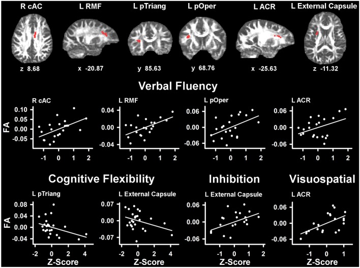
Diffusion tensor imaging (DTI) findings from emerging studies of cortical white-matter integrity in Parkinson's disease (PD) without dementia are inconclusive. When white-matter changes have been found, their relationship to cognitive functioning in PD has not been carefully investigated. To better characterize changes in tissue diffusivity and to understand their functional significance, the present study conducted DTI in 25 PD patients without dementia and 26 controls of similar ages. An automated tract-based DTI method was used. Fractional anisotropy (FA), mean diffusivity (MD), axial diffusivity (AD), and radial diffusivity (RD) were analyzed. Neuropsychological measures of executive functioning (working memory, verbal fluency, cognitive flexibility, inhibitory control) and visuospatial ability were then correlated with regions of interest that showed abnormal diffusivity in the PD group. We found widespread reductions in FA and increases in MD in the PD group relative to controls. These changes were predominantly related to an increase in RD. Increased AD in the PD group was limited to specific frontal tracks of the right hemisphere, possibly signifying more significant tissue changes. Motor symptom severity did not correlate with FA. However, different measures of executive functioning and visuospatial ability correlated with FA in different segments of tracts, which contain fiber pathways to cortical regions that are thought to support specific cognitive processes. The findings suggest that abnormal tissue diffusivity may be sensitive to subtle cognitive changes in PD, some of which may be prognostic of future cognitive decline.
Keywords: Parkinson’s disease; cerebral cortex; cognition; diffusion tensor imaging; white-matter.
Figures
Similar articles
-
Disruption of Inferior Longitudinal Fasciculus Microstructure in Parkinson's Disease: A Systematic Review of Diffusion Tensor Imaging Studies.Front Neurol. 2018 Jul 26;9:598. doi: 10.3389/fneur.2018.00598. eCollection 2018. Front Neurol. 2018. PMID: 30093877 Free PMC article. Review.
-
Relationship Between DTI Metrics and Cognitive Function in Alzheimer's Disease.Front Aging Neurosci. 2019 Jan 9;10:436. doi: 10.3389/fnagi.2018.00436. eCollection 2018. Front Aging Neurosci. 2019. PMID: 30687081 Free PMC article.
-
Progressive Decline in Gray and White Matter Integrity in de novo Parkinson's Disease: An Analysis of Longitudinal Parkinson Progression Markers Initiative Diffusion Tensor Imaging Data.Front Aging Neurosci. 2018 Oct 8;10:318. doi: 10.3389/fnagi.2018.00318. eCollection 2018. Front Aging Neurosci. 2018. PMID: 30349475 Free PMC article.
-
Multifocal alterations of white matter accompany the transition from normal cognition to dementia in Parkinson's disease patients.Brain Imaging Behav. 2019 Feb;13(1):232-240. doi: 10.1007/s11682-018-9863-7. Brain Imaging Behav. 2019. PMID: 29629498
-
The role of diffusion tensor imaging and fractional anisotropy in the evaluation of patients with idiopathic normal pressure hydrocephalus: a literature review.Neurosurg Focus. 2016 Sep;41(3):E12. doi: 10.3171/2016.6.FOCUS16192. Neurosurg Focus. 2016. PMID: 27581308 Review.
Cited by
-
Verbal Memory in Parkinson's Disease: A Combined DTI and fMRI Study.J Parkinsons Dis. 2015;5(4):793-804. doi: 10.3233/jpd-150623. J Parkinsons Dis. 2015. PMID: 27070003 Free PMC article.
-
Disruption of Inferior Longitudinal Fasciculus Microstructure in Parkinson's Disease: A Systematic Review of Diffusion Tensor Imaging Studies.Front Neurol. 2018 Jul 26;9:598. doi: 10.3389/fneur.2018.00598. eCollection 2018. Front Neurol. 2018. PMID: 30093877 Free PMC article. Review.
-
Effect of cerebral small vessel disease on cognitive impairment in Parkinson's disease: a systematic review and meta-analysis.Ann Transl Med. 2022 Mar;10(6):288. doi: 10.21037/atm-22-276. Ann Transl Med. 2022. PMID: 35433969 Free PMC article.
-
White matter abnormalities in the corpus callosum with cognitive impairment in Parkinson disease.Neurology. 2018 Dec 11;91(24):e2244-e2255. doi: 10.1212/WNL.0000000000006646. Epub 2018 Nov 14. Neurology. 2018. PMID: 30429273 Free PMC article.
-
Diffusion Measures of Subcortical Structures Using High-Field MRI.Brain Sci. 2023 Feb 24;13(3):391. doi: 10.3390/brainsci13030391. Brain Sci. 2023. PMID: 36979201 Free PMC article.
References
-
- Abrahams S., Goldstein L. H., Simmons A., Brammer M. J., Williams S. C., Giampietro V. P., et al. (2003). Functional magnetic resonance imaging of verbal fluency and confrontation naming using compressed image acquisition to permit overt responses. Hum. Brain Mapp. 20, 29–4010.1002/hbm.10126 - DOI - PMC - PubMed
Grants and funding
LinkOut - more resources
Full Text Sources
Other Literature Sources
Miscellaneous