Comparative Study
. 2013 Apr 22;8(4):e61597.
doi: 10.1371/journal.pone.0061597. Print 2013.

Efficacy of influenza vaccination and tamiflu® treatment--comparative studies with Eurasian Swine influenza viruses in pigs

Affiliations
Comparative Study

Efficacy of influenza vaccination and tamiflu® treatment--comparative studies with Eurasian Swine influenza viruses in pigs

Ralf Duerrwald et al. PLoS One. .

Abstract

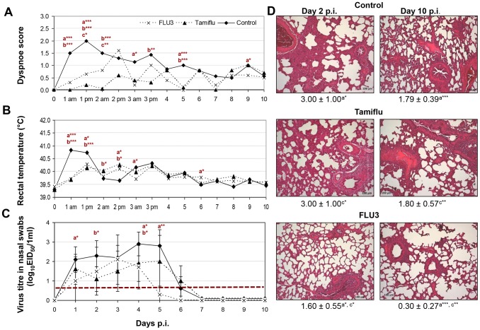
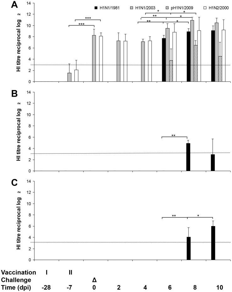
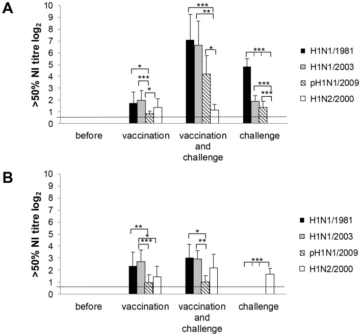
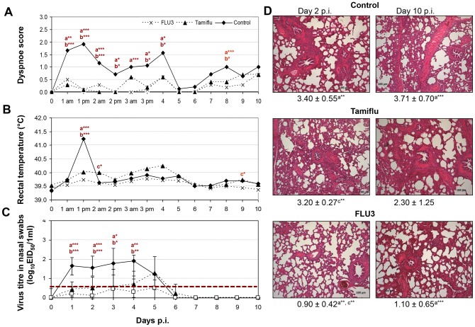
Recent epidemiological developments demonstrated that gene segments of swine influenza A viruses can account for antigenic changes as well as reduced drug susceptibility of pandemic influenza A viruses. This raises questions about the efficacy of preventive measures against swine influenza A viruses. Here, the protective effect of vaccination was compared with that of prophylactic Tamiflu® treatment against two Eurasian swine influenza A viruses. 11-week-old pigs were infected by aerosol nebulisation with high doses of influenza virus A/swine/Potsdam/15/1981 (H1N1/1981, heterologous challenge to H1N1 vaccine strain) and A/swine/Bakum/1832/2000 (H1N2/2000, homologous challenge to H1N2 vaccine strain) in two independent trials. In each trial (i) 10 pigs were vaccinated twice with a trivalent vaccine (RESPIPORC® FLU3; 28 and 7 days before infection), (ii) another 10 pigs received 150 mg/day of Tamiflu® for 5 days starting 12 h before infection, and (iii) 12 virus-infected pigs were left unvaccinated and untreated and served as controls. Both viruses replicated efficiently in porcine respiratory organs causing influenza with fever, dyspnoea, and pneumonia. Tamiflu® treatment as well as vaccination prevented clinical signs and significantly reduced virus shedding. Whereas after homologous challenge with H1N2/2000 no infectious virus in lung and hardly any lung inflammation were detected, the virus titre was not and the lung pathology was only partially reduced in H1N1/1981, heterologous challenged pigs. Tamiflu® application did not affect these study parameters. In conclusion, all tested preventive measures provided protection against disease. Vaccination additionally prevented virus replication and histopathological changes in the lung of homologous challenged pigs.


Conflict of interest statement

Competing Interests: The authors Ralf Duerrwald and Michael Schlegel are employed at the Research and Development department of IDT Biologika GmbH. The investigations reported in this manuscript were of scientific interest. The investigations could have been done by other institutions in a similar manner because the vaccine and drug used in the experiments are commercially available. The publication does not touch patents, consultancy or products in development. The vaccine administered is a marketed product of IDT Biologika GmbH. It was used because no other trivalent swine influenza vaccine containing H1N2 is licensed in Europe. This does not alter the authors' adherence to all PLOS ONE policies on sharing data and materials.

Figures

Figure 1
Figure 1. Protective effect of Tamiflu® in 11-week-old, A/swine/Potsdam/15/1981 (H1N1/1981) virus challenged pigs (n = 10) in comparison to RESPIPORC® FLU3-vaccinated (n = 10) and untreated animals (n = 12).
Dyspnoea (A), rectal temperatures (B), virus titres in nasal swabs, n = 10 animals/group/day until day 2 p.i. and n = 5 from day 3 p.i. on, exception: n = 12 untreated animals at day 0 to 2 p.i. and n = 7 untreated animals/day at 3 to 7 p.i. (C), and mean of histopathological scores with standard deviations and representative photographs of formalin fixed, HE stained lungs (D) are shown (am morning; pm afternoon; p statistical probability: *p<0.05, **p<0.01, ***p<0.001, a vaccinated group versus control group, b Tamiflu®-treated group versus control group, c vaccinated group versus Tamiflu®-treated group, Mann-Whitney-U-test). The detection limit of virus titre determination is shown as dotted line (C).
Figure 2
Figure 2. Influence of vaccination and Tamiflu® treatment on antibody kinetics in pigs challenged with A/swine/Potsdam/15/1981 (H1N1/1981) virus.
Hemagglutination inhibition assays were performed with serum from pigs vaccinated with RESPIPORC® FLU3 (A), pigs treated with Tamiflu® (B), and untreated pigs (C). Geometric mean and standard deviation of antibody titres determined in serum samples of 10 vaccinated and Tamiflu®-treated or 12 control animals/ /day are shown until day 2 p.i. and 5 or 7 from day 3–10 p.i., respectively. Mann-Whitney-U-test was used to calculate p statistical probability: *p<0.05, **p<0.01, ***p<0.001. Only significant differences are shown. The detection limit of HI antibody titre determination is shown as dotted line.
Figure 3
Figure 3. Neuraminidase inhibition by pig sera.
Sera were taken shortly before vaccination, 28 days before infection (“before”), 7 days after second vaccination (“vaccination”), 14 days after second vaccination and 7 days after infection with challenge virus (“vaccination and challenge”), and 7 days after infection with H1N1/1981 in the unvaccinated, untreated control group (“challenge”). Geometric mean of antibodies which cause 50% neuraminidase inhibition with standard deviations are shown (10 pigs before and at vaccination; 5 after vaccination and challenge, and 7 after challenge) for challenge with H1N1/1981 (A) and H1N2/2000 (B). Mann-Whitney-U-test was used to calculate p statistical probability: *p<0.05, **p<0.01, ***p<0.001. Statistics are shown for comparison within the groups (only statistical differences are shown). Data of statistical analysis for comparison between the groups (“before” versus “vaccination” and so on) are given in the text under results. The detection limit of NI antibody titre determination is shown as dotted line.
Figure 4
Figure 4. Antiviral activity of Tamiflu® in 11-week-old, A/swine/Bakum/1832/00 (H1N2/2000) virus infected pigs (n = 10) in comparison to RESPIPORC® FLU3-vaccinated (n = 10) and untreated animals (n = 12).
Dyspnoea (A), rectal temperatures (B), virus titres in nasal swabs, n = 10 animals/group/day until day 2 p.i. and n = 5 from day 3 p.i. on, exception: n = 12 untreated animals at day 0 to 2 p.i. and n = 7 untreated animals/day at 3 to 7 p.i. (C), and mean of histopathological scores with standard deviations and representative photographs of formalin fixed, HE stained lungs (D) are shown (am morning; pm afternoon; p statistical probability: *p<0.05, **p<0.01, ***p<0.001, a vaccinated group versus control group, b Tamiflu®-treated group versus control group, c vaccinated group versus Tamiflu®-treated group, Mann-Whitney-U-test). The detection limit of virus titre determination is shown as dotted line (C).
Figure 5
Figure 5. Influence of vaccination and Tamiflu® treatment on antibody kinetics in pigs challenged with A/swine/Bakum/1832/2000 (H1N2/2000) virus.
HI assay was carried out with serum from pigs vaccinated with RESPIPORC® FLU3 (A), pigs treated with Tamiflu® (B), and untreated pigs (C). Geometric mean and standard deviation of antibody titres determined in serum samples of 10 vaccinated and Tamiflu®-treated or 12 control animals/day are shown until day 2 p.i. and 5 or 7 from day 3–10 p.i., respectively. Mann-Whitney-U-test was used to calculate p statistical probability: *p<0.05, **p<0.01, ***p<0.001. The detection limit of HI antibody titre determination is shown as dotted line.

Similar articles

Cited by

References

    1. WHO (2009) Influenza (Seasonal).
    1. Palese P (2004) Influenza: old and new threats. Nat Med 10: S82–87. - PubMed
    1. Wright PF, Neumann G, Kawaoka Y (2007) Orthomyxoviruses. In: Knipe DM, Howley PM, editors. Fields Virology. Philadelphia, PA, 19106 USA: Lippincott Williams & Wilkins. pp. 1692–1740.
    1. Kuntz-Simon G, Madec F (2009) Genetic and antigenic evolution of swine influenza viruses in Europe and evaluation of their zoonotic potential. Zoonoses Public Health 56: 310–325. - PubMed
    1. Schrader C, Suess J (2003) Genetic characterization of a porcine H1N2 influenza virus strain isolated in Germany. Intervirology 46: 66–70. - PubMed

Publication types

MeSH terms