

Characterization of the non-polio enterovirus infections associated with acute flaccid paralysis in South-Western India
- PMID: 23630606
- PMCID: PMC3632520
- DOI: 10.1371/journal.pone.0061650
Characterization of the non-polio enterovirus infections associated with acute flaccid paralysis in South-Western India
Abstract
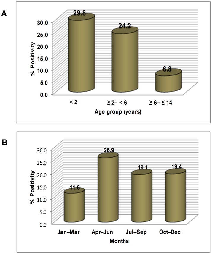
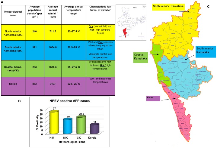
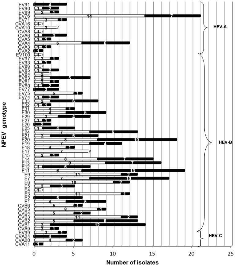
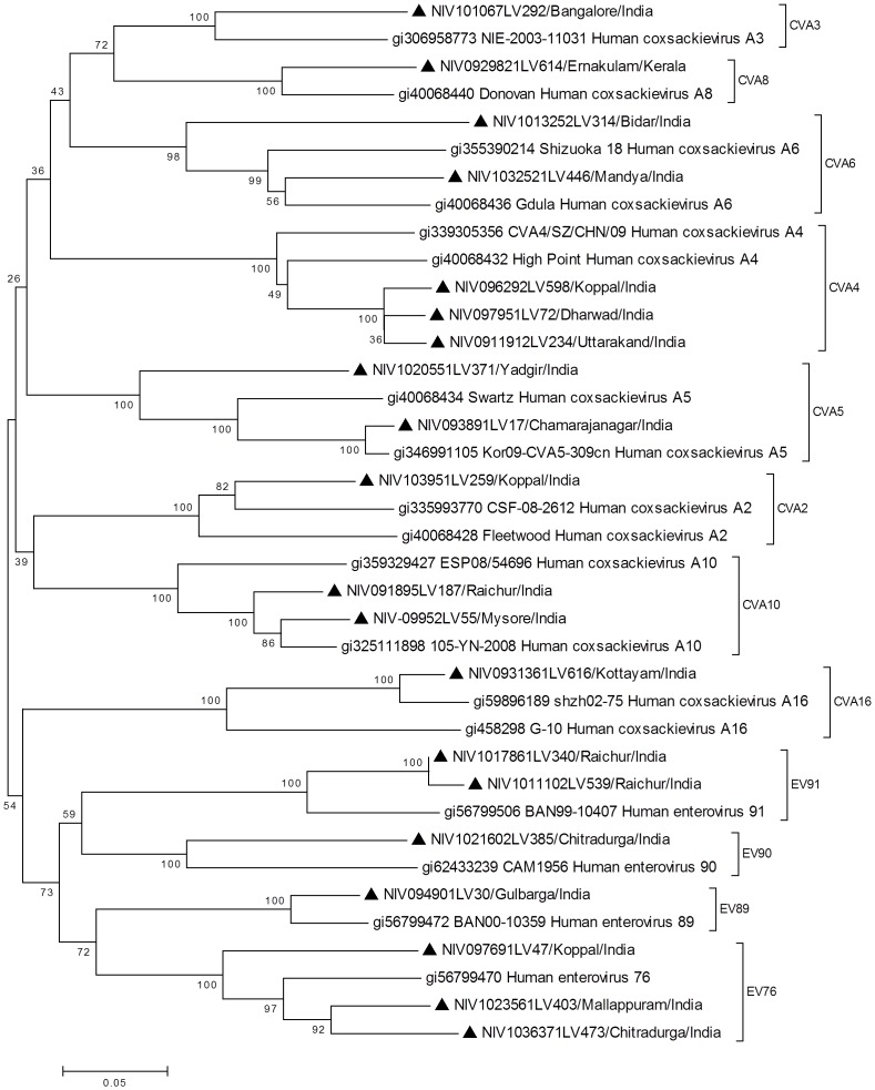
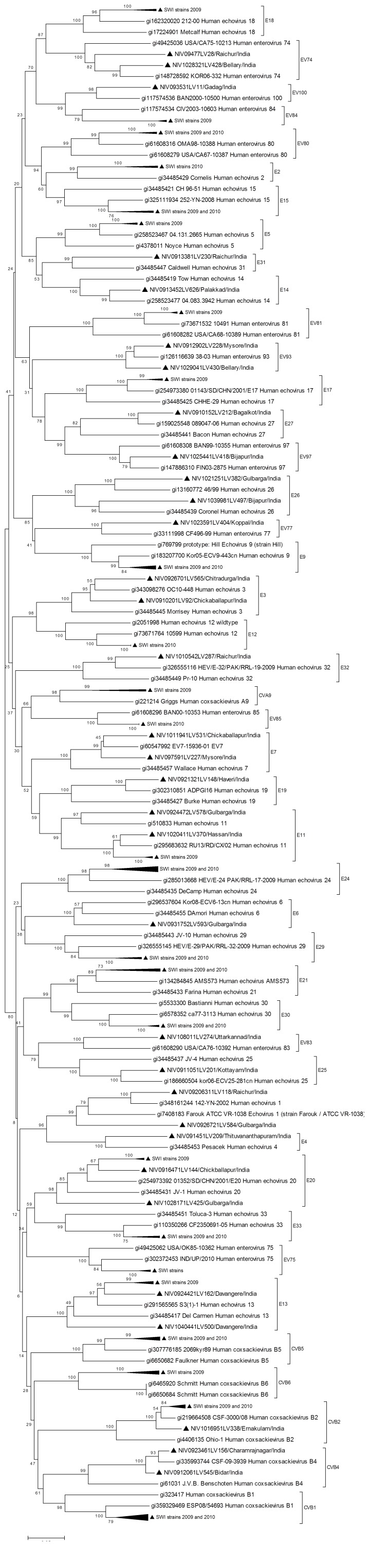
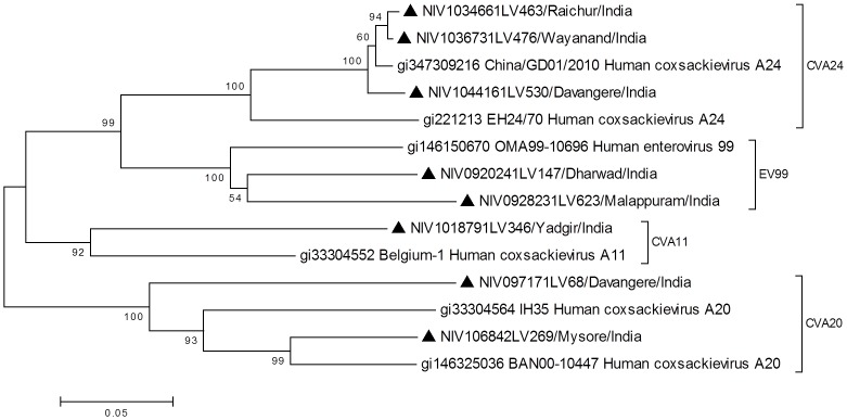
Non-polio enteroviruses (NPEVs) have been reported frequently in association with acute flaccid paralysis (AFP) cases during Polio Surveillance Programs (PSPs) worldwide. However, there is limited understanding on the attributes of their infections. This study reports characteristics of NPEVs isolated from AFP cases, investigated during PSPs held in 2009-2010, in Karnataka and Kerala states of south-western India having varied climatic conditions. NPEV cell culture isolates derived from stool specimens that were collected from 422 of 2186 AFP cases (<1-14 years age) and 17 of 41 asymptomatic contacts; and details of all AFP cases/contacts were obtained from National Polio Laboratory, Bangalore. The distribution of NPEV infections among AFP cases and circulation pattern of NPEV strains were determined by statistical analysis of the data. Genotyping of all NPEV isolates was carried out by partial VP1 gene sequencing and phylogenetic analysis. NPEV positive AFP cases were significantly higher in children aged <2 years; with residual paralysis; in summer months; and in regions with relatively hot climate. Genotyping of NPEVs identified predominance of human enteroviruses (HEV)-B species [81.9%-Echoviruses (E): 57.3%; coxsackieviruses (CV) B: 15%; numbered EVs: 8.9%; CVA9: 0.7%] and low levels of HEV-A [14.5%-CVA: 6%; numbered EVs: 8.5%] and HEV-C [3.6%-CVA: 2.6%; numbered EVs: 1%] species, encompassing 63 genotypes. EV76 (6.3%) and each of E3, CVB3 and E9 (4.97%) were found frequently during 2009 while E11 (6.7%), CVB1 (6.1%), E7 (5.1%) and E20 (5.1%) were detected commonly in 2010. A marked proportion of AFP cases from children aged <2 years; presenting with fever; and from north and south interior parts of Karnataka state was detected with E/numbered EVs than that found with CVA/CVB. This study highlights the extensive genetic diversity and diverse circulation patterns of NPEV strains in AFP cases from different populations and climatic conditions.
Conflict of interest statement
Figures
References
-
- Marx A, Glass JD, Sutter RW (2000) Differential diagnosis of acute flaccid paralysis and its role in poliomyelitis surveillance. Epidemiol Rev 22: 298–316. - PubMed
-
- Israeli E, Agmon-Levin N, Blank M, Chapman J, Shoenfeld Y (2012) Guillain-Barre syndrome–a classical autoimmune disease triggered by infection or vaccination. Clin Rev Allergy Immunol 42: 121–130. - PubMed
-
- Solomon T, Willison H (2003) Infectious causes of acute flaccid paralysis. Curr Opin Infect Dis 16: 375–381. - PubMed
-
- Kliegman R (2007) Nelson textbook of pediatrics: Saunders Elsevier Philadelphia.
-
- Pallansch MA, Roos R (2007) Enteroviruses. Polioviruses, Coxsackieviruses, Echoviruses and newer enteroviruses. In: Knipe DM, Howley PM, editors. Fields Virology. 5th ed. Lippincott Williams & Wilkin, PA, USA. pp. 839–893.
Publication types
MeSH terms
Substances
LinkOut - more resources
Full Text Sources
Other Literature Sources
Medical
