Review
. 2013 Aug;1296(1):11-30.
doi: 10.1111/nyas.12110. Epub 2013 Apr 30.

Combined neurostimulation and neuroimaging in cognitive neuroscience: past, present, and future

Affiliations
Free PMC article
Review

Combined neurostimulation and neuroimaging in cognitive neuroscience: past, present, and future

Sven Bestmann et al. Ann N Y Acad Sci. 2013 Aug.
Free PMC article

Abstract

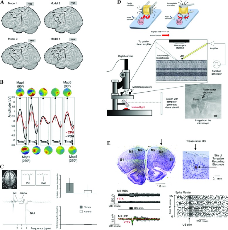
Modern neurostimulation approaches in humans provide controlled inputs into the operations of cortical regions, with highly specific behavioral consequences. This enables causal structure-function inferences, and in combination with neuroimaging, has provided novel insights into the basic mechanisms of action of neurostimulation on distributed networks. For example, more recent work has established the capacity of transcranial magnetic stimulation (TMS) to probe causal interregional influences, and their interaction with cognitive state changes. Combinations of neurostimulation and neuroimaging now face the challenge of integrating the known physiological effects of neurostimulation with theoretical and biological models of cognition, for example, when theoretical stalemates between opposing cognitive theories need to be resolved. This will be driven by novel developments, including biologically informed computational network analyses for predicting the impact of neurostimulation on brain networks, as well as novel neuroimaging and neurostimulation techniques. Such future developments may offer an expanded set of tools with which to investigate structure-function relationships, and to formulate and reconceptualize testable hypotheses about complex neural network interactions and their causal roles in cognition.

Keywords: EEG; MRS; causal inference; computational neurostimulation; effective connectivity; fMRI; state-dependence; transcranial magnetic stimulation.


Figures

Figure 1
Figure 1
Subcortical activity changes evoked by TMS as measured using fMRI and PET. (A) Brief bursts of rTMS to the left primary motor cortex (M1) evoke BOLD signal changes not only in the vicinity of the stimulation site but additionally in the ipsilateral motor thalamic nuclei (note: left and right reversal). These subcortical effects occur even during subthreshold TMS and therefore in the absence of evoked movements. (B) Changes in extracellular dopamine concentration measured in vivo using [(11)C]raclopride and positron emission tomography. Repetitive TMS of the left dorsolateral prefrontal cortex caused a reduction in [(11)C]raclopride binding in the left dorsal caudate nucleus compared with rTMS of the left occipital cortex. (C) Interaction of TMS (high and low intensity) and median nerve stimulation (ON and OFF) within the thalamus. BOLD signal in the thalamus was highest during combined right-hand somatosensory stimulation and high-intensity TMS over the right parietal cortex.
Figure 2
Figure 2
State-dependence of interhemispheric influences of TMS in the motor system. Short bursts of TMS (high vs. low intensity) were applied to the left PMd during left-hand isometric force production (or nonmotor rest), concurrently with fMRI. Within the task-related right PMd and M1 (i.e., contralateral to TMS, top), TMS at high intensity leads to a relative activity increase (bottom), compared to low-intensity control TMS. However, this effect is reversed during nonmotor rest, with high TMS now leading to a relative activity decrease in contralateral motor regions.
Figure 3
Figure 3
Anatomically remote effects of TMS to reveal the mechanisms of DLPFC-based control on WM representations. (A) Schematic of the right DLPFC stimulation site (upper), a region involved in distractor mitigation during WM. The impact of stimulation was assessed in posterior visual category-specific areas (lower; fusiform face area (FFA) in red, parahippocampal place area (PPA) in blue)). (B) Interparticipant mean BOLD percent signal changes due to effective vs. ineffective DLPFC-TMS are shown in FFA (upper) and PPA (lower). Effective TMS increased BOLD in FFA specifically when faces were memory targets, in the presence of house distractors. Analogously, effective TMS increased activity in PPA when houses were memory targets and faces were distractors. Thus, DLPFC stimulation has an impact on the posterior region representing the current memory target (rather than the current distractor), but only in the presence of distraction.
Figure 4
Figure 4
Effects of intraparietal TMS during perceptual decision making on sensorimotor oscillatory activity. (A) TMS-induced change in psychometric function during a perceptual decision-making task, for the left medial intraparietal area (MIP; green) vs. V5 (red) control stimulation (B) revealed a specific correlation between TMS-induced behavioral changes during a perceptual decision task, and concurrently measured β-band changes for electrode positions overlying sensorimotor cortex in both hemispheres (C).
Figure 5
Figure 5
The future of concurrent neurostimulation and neuroimaging in humans for studies of cognition. (A) Network analyses such as dynamic causal modeling provide intermediate levels of description that link the physiological impact of neurostimulation and its behavioral consequences, and allow for generating testable hypotheses about resulting network connectivity changes. Adapted from Li et al. with permission. (B) Neurostimulation-induced entrainment of intrinsic cortical rhythms can have direct consequences for perception, depending on the frequency of stimulation. This allows for identifying the causal role of oscillatory activity for behavior, and holds the possibility to shape behavior through the selective entrainment of local and distributed oscillatory activity. Adapted from Thut et al. with permission. (C) Combined magnetic resonance spectroscopy can quantify the specific neurotransmitter changes induced by neurostimulation, potentially including online neurostimulation protocols. Moreover, the changes can be directly related to behavior, and thereby provide causal links between stimulation-evoked neurotransmitter changes in focal and defined parts of the brain, and cognition. Adapted from Stagg et al. with permission. (D) Micro-stimulation coils in principle allow for selective stimulation of cortical micro-circuits, and include the possibility for application during neuroimaging Adapted from Bonmassar et al. with permission. (E) Ultrasound stimulation holds promise to allow for targeted and selective stimulation of neural tissue throughout the brain, including subcortical structures. Adapted from Tufail et al. with permission. Note that for the examples in D–E, demonstration of their applicability in humans is pending.

References

    1. Nitsche MA, Paulus W. Excitability changes induced in the human motor cortex by weak transcranial direct current stimulation. J. Physiol. 2000;527:633–639. - PMC - PubMed
    1. Nitsche MA, Paulus W. Transcranial direct current stimulation—update 2011. Restor. Neurol. Neurosci. 2011;29:463–492. - PubMed
    1. Stagg CJ, Nitsche MA. Physiological basis of transcranial direct current stimulation. Neuroscientist. 2011;17:37–53. - PubMed
    1. Holland R, Leff AP, Josephs O, et al. Speech facilitation by left inferior frontal cortex stimulation. Curr. Biol. 2011;21:1403–1407. - PMC - PubMed
    1. Antal A, Polania R, Schmidt-Samoa C, et al. Transcranial direct current stimulation over the primary motor cortex during fMRI. Neuroimage. 2011;55:590–596. - PubMed

Publication types