

Phosphatidylinositol-4,5-bisphosphate is enriched in granulovacuolar degeneration bodies and neurofibrillary tangles
- PMID: 23631697
- PMCID: PMC4298759
- DOI: 10.1111/nan.12056
Phosphatidylinositol-4,5-bisphosphate is enriched in granulovacuolar degeneration bodies and neurofibrillary tangles
Abstract
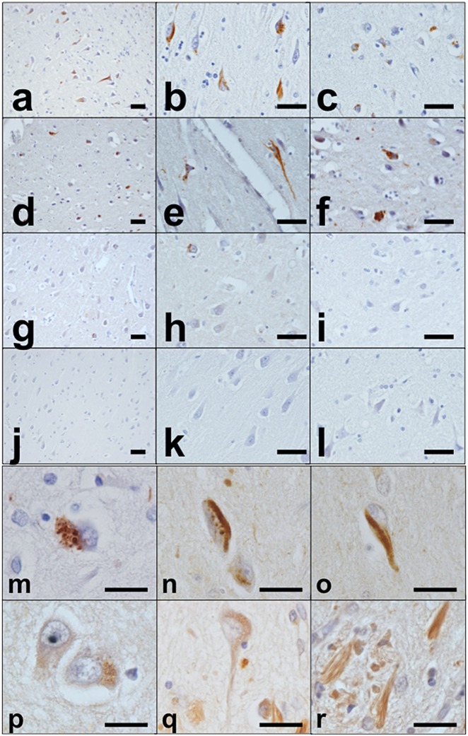
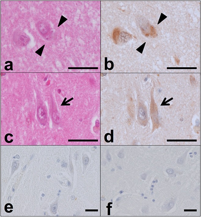
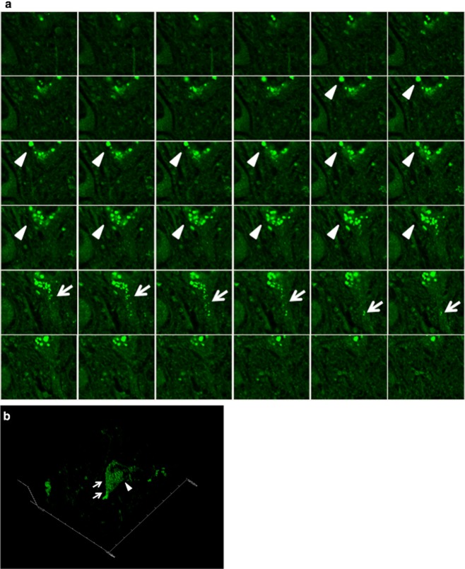
Aims: Among the pathological findings in Alzheimer's disease (AD), the temporal and spatial profiles of granulovacuolar degeneration (GVD) bodies are characteristic in that they seem to be related to those of neurofibrillary tangles (NFTs), suggesting a common mechanism underlying the pathogenesis of these structures. Flotillin-1, a marker of lipid rafts, accumulates in lysosomes of tangle-bearing neurones in AD patients. In addition, recent reports have shown that GVD bodies accumulate at the nexus of the autophagic and endocytic pathways. The aim of this study was to elucidate the distribution of the lipid component of lipid rafts, phosphatidylinositol-4,5-bisphosphate [PtdIns(4,5)P2], in AD and other neurodegenerative disorders.
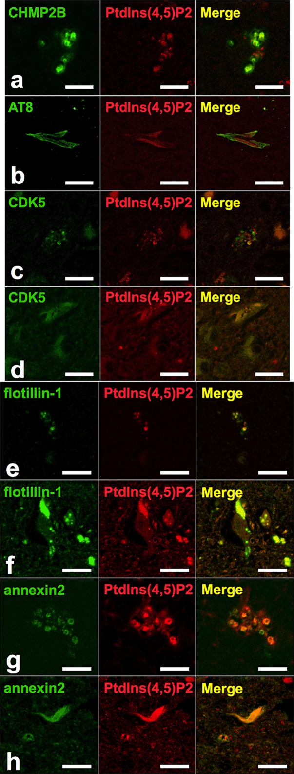
Methods: We compared PtdIns(4,5)P2 immunoreactivity in the hippocampus, entorhinal cortex and neocortex of five AD cases, 17 cases of other neurodegenerative disorders and four controls. In addition, we performed double staining using markers of GVD, NFTs and lipid rafts for further characterization.
Results: Immunohistochemical analysis revealed that PtdIns(4,5)P2 was selectively enriched in GVD bodies and NFTs. Although immunoreactivity for PtdIns(4,5)P2 was also evident in NFTs composed of hyperphosphorylated tau, PtdIns(4,5)P2 was segregated from phosphorylated tau within NFTs by double immunofluorescence staining. In contrast, PtdIns(4,5)P2 colocalized with the lipid raft markers flotillin-1 and annexin 2, within GVD bodies and NFTs.
Conclusions: These results suggest that lipid raft components including PtdIns(4,5)P2 play a role in the formation of both GVD bodies and NFTs.
Keywords: 5-bisphosphate; Alzheimer's disease; granulovacuolar degeneration; lipid raft; neurofibrillary tangle; phosphatidylinositol-4.
© 2013 The Authors. Neuropathology and Applied Neurobiology published by John Wiley & Sons Ltd. on behalf of British Neuropathological Society.
Figures
References
-
- Kadokura A, Yamazaki T, Kakuda S, Makioka K, Lemere CA, Fujita Y, Takatama M, Okamoto K. Phosphorylation-dependent TDP-43 antibody detects intraneuronal dot-like structures showing morphological characters of granulovacuolar degeneration. Neurosci Lett. 2009;463:87–92. - PubMed
-
- Baig S, van Helmond Z, Love S. Tau hyperphosphorylation affects Smad 2/3 translocation. Neuroscience. 2009;163:561–570. - PubMed
-
- Yamazaki Y, Takahashi T, Hiji M, Kurashige T, Izumi Y, Yamawaki T, Matsumoto M. Immunopositivity for ESCRT-III subunit CHMP2B in granulovacuolar degeneration of neurons in the Alzheimer’s disease hippocampus. Neurosci Lett. 2010;477:86–90. - PubMed
-
- Leroy K, Boutajangout A, Authelet M, Woodgett JR, Anderton BH, Brion JP. The active form of glycogen synthase kinase-3beta is associated with granulovacuolar degeneration in neurons in Alzheimer’s disease. Acta Neuropathol. 2002;103:91–99. - PubMed
-
- Nakamori M, Takahashi T, Yamazaki Y, Kurashige T, Yamawaki T, Matsumoto M. Cyclin-dependent kinase 5 immunoreactivity for granulovacuolar degeneration. Neuroreport. 2012;23:867–872. - PubMed
Publication types
MeSH terms
Substances
LinkOut - more resources
Full Text Sources
Other Literature Sources
Medical
