. 2013 Apr 30:6:20.
doi: 10.1186/1756-6606-6-20.

Abnormal interneuron development in disrupted-in-schizophrenia-1 L100P mutant mice

Affiliations

Abnormal interneuron development in disrupted-in-schizophrenia-1 L100P mutant mice

Frankie H F Lee et al. Mol Brain. .

Abstract

Background: Interneuron deficits are one of the most consistent findings in post-mortem studies of schizophrenia patients and are likely important in the cognitive deficits associated with schizophrenia. Disrupted-in-Schizophrenia 1 (DISC1), a strong susceptibility gene for schizophrenia and other mental illnesses, is involved in neurodevelopment, including that of interneurons. However, the mechanism by which DISC1 regulates interneuron development remains unknown. In this study, we analyzed interneuron histology in the Disc1-L100P single point mutation mouse, that was previously shown to have behavioral abnormalities and cortical developmental defects related to schizophrenia.

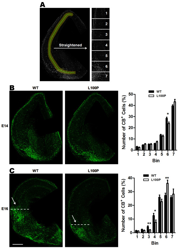
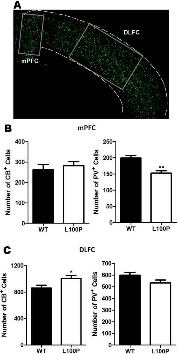
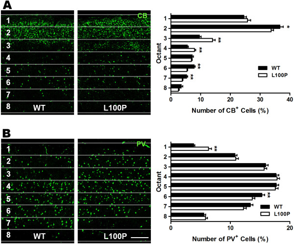
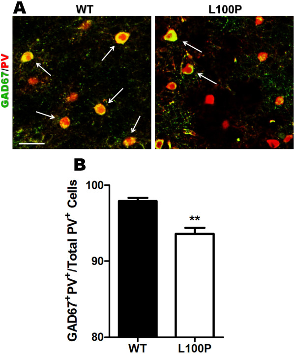
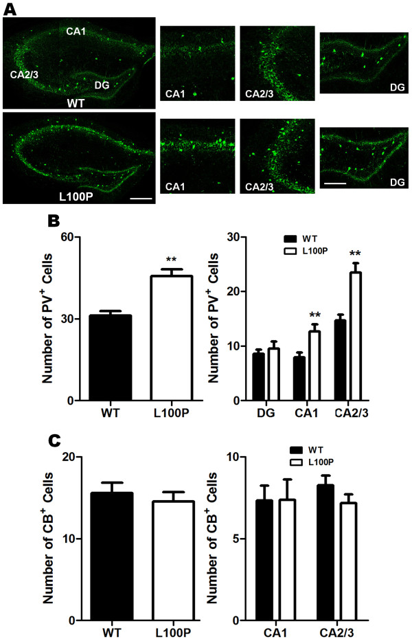
Results: We sought to determine whether a Disc1-L100P point mutation in the mouse would alter interneuron density and location. First, we examined interneuron position in the developing mouse cortex during embryonic days 14-16 as an indicator of interneuron tangential migration, and found striking migration deficits in Disc1-L100P mutants. Further analysis of adult brains revealed that the Disc1-L100P mutants have selective alterations of calbindin- and parvalbumin-expressing interneurons in the cortex and hippocampus, decreased GAD67/PV co-localization and mis-positioned interneurons across the neocortex when compared to wild-type littermates.

Conclusion: Our results are consistent with the anomalies seen in post-mortem schizophrenia studies and other Disc1 mutant mouse models. Future research is required to determine the specific mechanisms underlying these cellular deficits. Overall, these findings provide further evidence that DISC1 participates in interneuron development and add to our understanding of how DISC1 variants can affect susceptibility to psychiatric illness.


Figures

Figure 1
Figure 1
Disrupted interneuron tangential migration in Disc1-L100P embryonic mice. (A) Coronal sections of either E14 or E16 embryonic brains immunostained with CB were straightened and divided into seven equidistant bins covering the tangential migratory pathway of interneurons. Fluorescently-labeled cell numbers were counted in each bin and normalized to the total number in all bins. The distribution of these interneurons was significantly different for both E14 and E16 time points between WT and Disc1-L100P (two-way ANOVA, p < 0.01). (B) E14 CB-immunolabeled images of WT and Disc1-L100P mice are shown on the left. Disc1-L100P mice had a significantly lower proportion of CB-cells in the ventral bin 6 when compared to WT controls (n = 25–28 sections from 4 different embryos per group, t-test, p < 0.05). (C) At E16, a more pronounced difference in interneuron tangential migration was observed between WT and Disc1-L100P mice (white arrows). Disc1-L100P mutants had significantly less CB+ cells in bins 3 and 4, while more remained near the ventral bin 6 when compared to WT (n = 19–24 sections from 4 different embryos per group, t-test, p < 0.01). Scale Bar, 300 μm. All data are shown as mean ± SEM; * p < 0.05, ** p < 0.01 versus WT. CB, calbindin.
Figure 2
Figure 2
Differential alterations in CB- and PV-labeled interneurons in the mPFC and DLFC of Disc1-L100P adult mice. (A) A representative section used for counting CB- and PV-labeled cell densities is shown with boxes depicting the areas analyzed for the mPFC and DLFC regions. Scale Bar, 500 μm. (B) Quantification of CB+ and PV+ cells in the mPFC showed significantly fewer PV-interneurons in the Disc1-L100P mutants but no significant difference with CB-interneurons when compared to WT (n = 13–14 from 4 mice per group; t-test, p < 0.01). (C) In contrast, Disc1-L100P mutants have significantly more CB+ cells but not PV+ cells in the DLFC versus WT (n = 21–22 from 4 mice per group, t-test, p < 0.05). All data are shown as mean ± SEM; * p < 0.05, ** p < 0.01 versus WT. CB, calbindin; DLFC, dorsal lateral frontal cortex; mPFC, medial prefrontal cortex; PV, parvalbumin.
Figure 3
Figure 3
Altered cortical interneuron laminar distribution in Disc1-L100P adult mice. Quantification of both CB- and PV-labeled cells was achieved by counting the number of fluorescent cells in each octant and expressed as a percentage of total number per ROI of fixed size across the neocortex. (A) There was a significant interaction effect with CB-interneurons across the octants between WT and Disc1-L100P mice (two-way ANOVA, p < 0.01) with proportionally more CB-interneurons in the middle cortical layers of octants 3 and 4 (t-test, p < 0.01). Conversely, there were fewer CB+ cells in the second, sixth and seventh octants in the Disc1-L100P mutant versus WT (n = 52 ROIs from 4 different mice per group; t-test, p < 0.01). (B) PV-labeled interneurons were significantly different in their distribution across the octants between WT and Disc1-L100P mutants (two-way ANOVA, p < 0.01). More PV + cells were observed in WT of octant 6 but less in superficial cortical layers of octant 1 than in the mutants (n = 84–90 ROIs from 5 different mice per group; t-test, p < 0.01). Scale Bar, 300 μm. All data are shown as mean ± SEM; * p < 0.05, ** p < 0.01 versus WT. CB, calbindin; PV, parvalbumin; ROI, region of interest.
Figure 4
Figure 4
Aberrant tangential distribution of PV-interneurons in Disc1-L100P adult mice. (A) Four sampling ROIs immunolabeled with either CB or PV antibodies were outlined across the neocortex along the medial-lateral axis. The number of CB+ and PV+ cells were counted and expressed as a percentage of total cells in each ROI. (B) There was no significant difference in the tangential distribution of CB+ cells between WT and Disc1-L100P mice (n = 12–13 ROIs from 4 different mice per group). (C) PV-labeled cells were distributed differently depending on genotype. The Disc1-L100P mutants had a significantly higher proportion of PV+ cells located laterally (bin 1) but fewer PV+ cells in medial bin 4 when compared to WT (n = 22 ROIs from 4 different mice per group; two-way ANOVA, t-test, p < 0.05). Scale Bar, 500 μm. All data are shown as mean ± SEM; * p < 0.05 versus WT. CB, calbindin; PV, parvalbumin; ROI, region of interest.
Figure 5
Figure 5
Reduced GAD67/PV co-localization in Disc1-L100P adult mutants. (A) GAD67 (green) and PV (red) fluorescently labeled images in WT and Disc1-L100P mutants. White arrows indicate those with more than 50% overlap of GAD67 and PV. Scale Bar, 50 μm. (B) Quantification of the percentage of GAD67+PV+ cells per total PV+ cells revealed significantly less GAD67 expression within PV-cells in Disc1-L100P mice compared to WT (n = 106–128 ROIs from 4 different mice per group). All data are shown as mean ± SEM; ** p < 0.01 versus WT. GAD67, glutamic acid decarboxylase (67 kDa); PV, parvalbumin.
Figure 6
Figure 6
Selective increase of PV-interneurons in CA1 and CA2/3 regions of the hippocampus in Disc1-L100P adult mutant mice. (A) PV-immunostained images of the hippocampus in WT and Disc1-L100P mice were divided into CA1, CA2/3 and DG subfields in which labeled cells were counted. Scale Bar, 300 μm. A higher magnification of each subfield is shown on the right. Scale Bar, 100 μm. (B) Disc1-L100P mutants had significantly more PV-interneurons in the hippocampus, particularly within CA1 and CA2/3 subfields when compared to WT (n = 12 ROIs for each region from 4 different mice per group). (C) There was no significant difference in the number of CB+ interneurons within the CA1 and CA2/3 regions between WT and Disc1-L100P mutants. DG was excluded from analysis due to the absence of CB-interneurons. All data are shown as mean ± SEM; ** p < 0.01 versus WT. CB, calbindin; PV, parvalbumin; CA, Cornu Ammonis; DG, dentate gyrus.

References

    1. Miller EK, Cohen JD. An integrative theory of prefrontal cortex function. Annu Rev Neurosci. 2001;24:167–202. doi: 10.1146/annurev.neuro.24.1.167. - DOI - PubMed
    1. Marin O. Interneuron dysfunction in psychiatric disorders. Nat Rev Neurosci. 2012;13:107–120. - PubMed
    1. Gonzalez-Burgos G, Hashimoto T, Lewis DA. Alterations of cortical GABA neurons and network oscillations in schizophrenia. Curr Psychiatry Rep. 2010;12:335–344. doi: 10.1007/s11920-010-0124-8. - DOI - PMC - PubMed
    1. Blum BP, Mann JJ. The GABAergic system in schizophrenia. Int J Neuropsychopharmacol. 2002;5:159–179. - PubMed
    1. Lewis DA, Hashimoto T, Volk DW. Cortical inhibitory neurons and schizophrenia. Nat Rev Neurosci. 2005;6:312–324. doi: 10.1038/nrn1648. - DOI - PubMed

Publication types

MeSH terms

LinkOut - more resources