. 2013 Apr 30;4(3):e00018-13.
doi: 10.1128/mBio.00018-13.

STING-dependent recognition of cyclic di-AMP mediates type I interferon responses during Chlamydia trachomatis infection

Affiliations

STING-dependent recognition of cyclic di-AMP mediates type I interferon responses during Chlamydia trachomatis infection

Jeffrey R Barker et al. mBio. .

Abstract

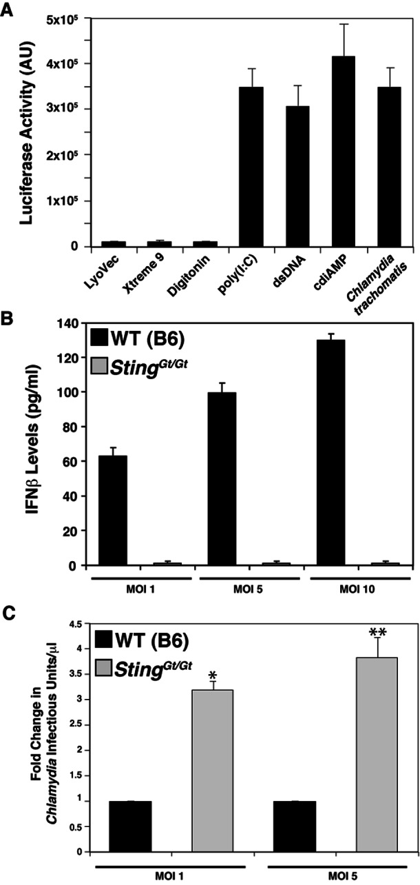
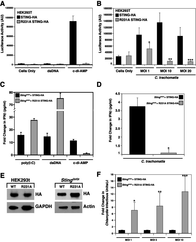
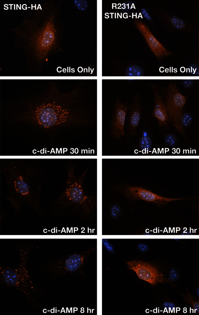
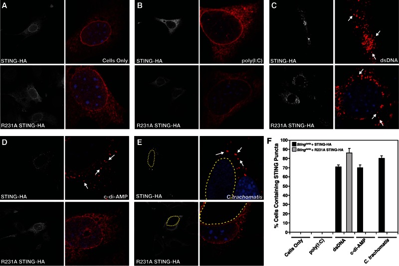
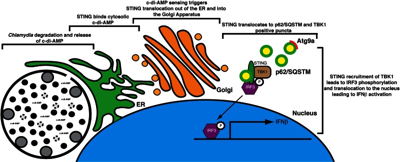
STING (stimulator of interferon [IFN] genes) initiates type I IFN responses in mammalian cells through the detection of microbial nucleic acids. The membrane-bound obligate intracellular bacterium Chlamydia trachomatis induces a STING-dependent type I IFN response in infected cells, yet the IFN-inducing ligand remains unknown. In this report, we provide evidence that Chlamydia synthesizes cyclic di-AMP (c-di-AMP), a nucleic acid metabolite not previously identified in Gram-negative bacteria, and that this metabolite is a prominent ligand for STING-mediated activation of IFN responses during infection. We used primary mouse lung fibroblasts and HEK293T cells to compare IFN-β responses to Chlamydia infection, c-di-AMP, and other type I IFN-inducing stimuli. Chlamydia infection and c-di-AMP treatment induced type I IFN responses in cells expressing STING but not in cells expressing STING variants that cannot sense cyclic dinucleotides but still respond to cytoplasmic DNA. The failure to induce a type I IFN response to Chlamydia and c-di-AMP correlated with the inability of STING to relocalize from the endoplasmic reticulum to cytoplasmic punctate signaling complexes required for IFN activation. We conclude that Chlamydia induces STING-mediated IFN responses through the detection of c-di-AMP in the host cell cytosol and propose that c-di-AMP is the ligand predominantly responsible for inducing such a response in Chlamydia-infected cells.

Importance: This study shows that the Gram-negative obligate pathogen Chlamydia trachomatis, a major cause of pelvic inflammatory disease and infertility, synthesizes cyclic di-AMP (c-di-AMP), a nucleic acid metabolite that thus far has been described only in Gram-positive bacteria. We further provide evidence that the host cell employs an endoplasmic reticulum (ER)-localized cytoplasmic sensor, STING (stimulator of interferon [IFN] genes), to detect c-di-AMP synthesized by Chlamydia and induce a protective IFN response. This detection occurs even though Chlamydia is confined to a membrane-bound vacuole. This raises the possibility that the ER, an organelle that innervates the entire cytoplasm, is equipped with pattern recognition receptors that can directly survey membrane-bound pathogen-containing vacuoles for leaking microbe-specific metabolites to mount type I IFN responses required to control microbial infections.


Figures

FIG 1
FIG 1
The C. trachomatis protein DacA (CT102) displays DAC activity. (A) Domain architecture of B. subtilis DAC domain-containing proteins DisA, YojJ, and Ybbp with L. monocytogenes DacA and the C. trachomatis putative DAC CT012. HhH1, helix-hairpin-helix motif; TM, three TM domains). (B) Amino acid sequence alignment of conserved domain regions of C. trachomatis DacA and DACs from c-di-AMP-producing bacteria with conserved residues critical for c-di-AMP synthesis indicated (asterisks). One hundred percent similarity, black; 80 to 100% similarity, dark gray; 60 to 80% similarity, light gray; <60% similarity, white. (C) Schematics of three C. trachomatis DacA recombinant variants used in this work to monitor c-di-AMP synthesis in E. coli. (D) UPLC-based quantification of c-di-AMP levels from nucleotide extracts of E. coli expressing full-length Chlamydia DacA, the DAC domain of DacA, or DacA with a D164N point mutation in the putative active site with corresponding immunoblot assays showing levels of DacA in soluble protein extracts. The data shown are means ± standard deviations of two independent experiments. Commercial c-di-AMP (Biolog) was used for calibration and quantification of nucleotide extracts.
FIG 2
FIG 2
c-di-AMP is present in nucleic acid extracts from cells infected with C. trachomatis. (A) c-di-AMP levels in extracts of HeLa cells infected with C. trachomatis were quantified by UPLC at the indicated time points. The corresponding immunoblot shows Chlamydia DacA and MOMP expression levels. Asterisks indicate monomeric (*) and dimeric (**) forms of DacA. (B) MEFs induce type I IFN in response to Chlamydia infection. MEFs stably expressing an ISRE fused to a luciferase reporter gene were infected with C. trachomatis for the indicated times and lysed, and luciferase levels were assessed by an enzymatic assay. AU, arbitrary units. (C) c-di-AMP levels in lysates of density gradient-purified RBs and EBs. The signal for c-di-AMP was sensitive to SVP. The data shown are means ± standard deviations of two technical replicates.
FIG 3
FIG 3
STING is required for the expression of type I IFN in mouse MLFs during C. trachomatis infection. (A) MLFs induce type I IFNs in response to various microbial-signal-like signals. MLFs were transfected with dsDNA or poly(I-C), loaded with c-di-AMP by digitonin permeabilization, or infected with C. trachomatis for 30 h. Cells treated with transfection (LuoVec and Xtreme9) or cell permeabilization reagents alone were included as controls. Levels of secreted type I IFNs in cell supernatants were assessed with a reporter cell line stably expressing an ISRE fused to a luciferase reporter gene and monitoring of luciferase enzymatic activity. AU, arbitrary units. (B) IFN-β responses to C. trachomatis infection requires STING. MLFs from wild-type (WT) and STINGGt/Gt (Goldenticket) mice were infected with C. trachomatis at the indicated MOIs for 30 h, and the levels of IFN-β in cell supernatants was determined by ELISA. (C) STING-deficient (STINGGt/Gt) MLFs are permissive for C. trachomatis replication. Wild-type and STINGGt/Gt MLFs were infected with C. trachomatis at the indicated MOIs, and the yield of infectious units was determined at 36 hpi. Fold change represents the ratio of the number of infectious units/μl observed in Goldenticket fibroblasts to that observed in wild-type fibroblasts. All data are means ± standard deviations of three independent samples ×, P < 0.001; ∗∗, P <0.001 (one-way analysis of variance and Newman-Keuls multiple-comparison test).
FIG 4
FIG 4
c-di-AMP is the main ligand responsible for Chlamydia-mediated type I IFN responses. (A and B) STING expression is sufficient for the induction of IFN responses during C. trachomatis infection. HEK293T cells stably expressing an ISRE-luciferase reporter were transduced with a wild-type STING-HA (black) or a STINGR231A-HA (gray) retroviral vector and treated with dsDNA or c-di-AMP (A) or infected with C. trachomatis for 30 h at the MOIs indicated (B). ×, P < 0.05; **, P < 0.001; ***, P < 0.001 (one-way analysis of variance and Newman-Keuls multiple-comparison test). Cells expressing mutant STINGR231A cannot induce type I IFNs in response to c-di-AMP. AU, arbitrary units. (C to F) MLFs expressing STING variants that cannot signal in response to cyclic dinucleotides fail to induce type I IFNs during C. trachomatis infection. Goldenticket MLFs transduced with a wild-type STING-HA (black) or a STINGR231A-HA (gray) construct were treated with poly(I-C), dsDNA, or c-di-AMP (C) or infected with C. trachomatis for 30 h (D). *, P < 0.05 (one-way analysis of variance and Newman-Keuls multiple-comparison test). Supernatants were tested for IFN-β levels by ELISA. Fold changes represent the ratios of total IFN-β detected by ELISA (in pg/ml) in cells treated with stimulants to that in untreated cells (C) and that in infected cells to that in uninfected cells (D). (E) Immunoblot analysis with anti-HA (STING) antibody of protein lysates from HEK293T cells and Goldenticket MLFs expressing various STING constructs indicates that comparable levels of STING were expressed in these cell lines. Glyceraldehyde 3-phosphate dehydrogenase (GAPDH) and actin served as loading controls. WT, wild type. (F) Goldenticket MLFs expressing wild-type STING-HA (black) or STINGR231A-HA were infected with C. trachomatis, and infectious units were assessed as described in the legend to Fig. 3C. Note that Goldenticket MLF expressing the STING variant that cannot recognize ci-di-AMP did not induce IFN-β and control C. trachomatis replication. *, P < 0.05; **, P < 0.05; ***, P < 0.001 (one-way analysis of variance and Newman-Keuls multiple-comparison test). Fold changes represent the ratios of infectious units/μl observed in Goldenticket MLF complemented with STING-HAR231A-HA to those in wild-type STING-HA. The data shown are means ± standard deviations of three independent samples.
FIG 5
FIG 5
STING translocates to cytoplasmic structures in response to c-di-AMP. Goldenticket MLFs transduced with wild-type STING-HA (top) or STINGR231A-HA (bottom) were treated with c-di-AMP for the indicated times. Cells were immunostained with anti-HA (STING) antibody and imaged by epifluorescence microscopy.
FIG 6
FIG 6
(A) A STING variant (STINGR231A) that does not signal in response to c-di-AMP fails to translocate to cytoplasmic complexes upon Chlamydia infection. Immortalized Goldenticket MLFs expressing a wild-type STING-HA or a cyclic dinucleotide-blind STINGR231A-HA construct were treated with digitonin alone (A), transfected with the dsRNA analogue poly(I-C) (B) or dsDNA (C) for 24 h, loaded with c-di-AMP after digitonin permeabilization for 8 h (D), or infected with C. trachomatis for 30 h (E). Cells were fixed and stained with anti-HA (STING) antibody and imaged by confocal laser scanning microscopy. Note that STINGR231A cannot translocate to cytoplasmic punctate structures (arrows) in response to C. trachomatis infection or c-di-AMP stimulation but is responsive to dsDNA treatment. (F) Quantification of the percentage of cells displaying STING in cytoplasmic puncta after treatment with various inducers of type I IFNs. Cells displaying puncta (arrows) were scored as positive for STING translocation (n = 150).
FIG 7
FIG 7
Model of activation of STING-dependent IFN-β responses in Chlamydia-infected cells. The bacterial metabolite c-di-AMP is synthesized by Chlamydia and sensed by STING at ER membranes enveloping the inclusion. STING is transported out of the ER (26) to the Golgi apparatus and eventually to p62/SQSTSM cytoplasmic complexes (see Fig. S3 in the supplemental material). As previously shown, the recruitment of TBK1 and IRF3 to these signaling platforms leads to IRF3 phosphorylation and translocation to the nucleus and expression of type I IFN genes (29, 71).

References

    1. Haggerty CL, Gottlieb SL, Taylor BD, Low N, Xu F, Ness RB. 2010. Risk of sequelae after Chlamydia trachomatis genital infection in women. J. Infect. Dis. 201:S134–S155 - PubMed
    1. Abdelrahman YM, Belland RJ. 2005. The chlamydial developmental cycle. FEMS Microbiol. Rev. 29:949–959 - PubMed
    1. Hybiske K, Stephens RS. 2007. Mechanisms of host cell exit by the intracellular bacterium chlamydia. Proc. Natl. Acad. Sci. U. S. A. 104:11430–11435 - PMC - PubMed
    1. Takeuchi O, Akira S. 2010. Pattern recognition receptors and inflammation. Cell 140:805–820 - PubMed
    1. Monroe KM, McWhirter SM, Vance RE. 2010. Induction of type I interferons by bacteria. Cell. Microbiol. 12:881–890 - PMC - PubMed

Publication types