Review
. 2013 Jun 14;288(24):17074-81.
doi: 10.1074/jbc.R113.473108. Epub 2013 Apr 30.

Reactive intermediates in cytochrome p450 catalysis

Affiliations
Review

Reactive intermediates in cytochrome p450 catalysis

Courtney M Krest et al. J Biol Chem. .

Abstract

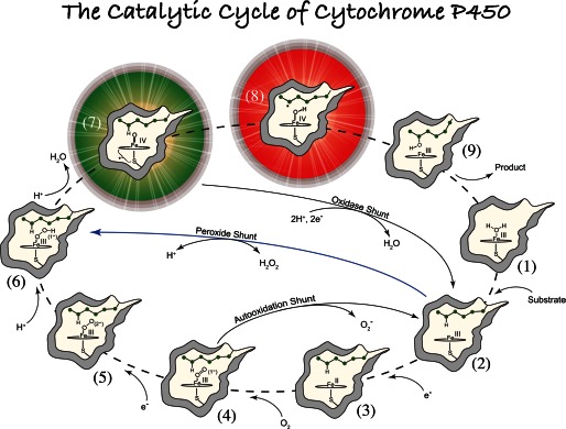
Recently, we reported the spectroscopic and kinetic characterizations of cytochrome P450 compound I in CYP119A1, effectively closing the catalytic cycle of cytochrome P450-mediated hydroxylations. In this minireview, we focus on the developments that made this breakthrough possible. We examine the importance of enzyme purification in the quest for reactive intermediates and report the preparation of compound I in a second P450 (P450ST). In an effort to bring clarity to the field, we also examine the validity of controversial reports claiming the production of P450 compound I through the use of peroxynitrite and laser flash photolysis.

Keywords: Cytochrome P450; Enzyme Catalysis; Enzyme Purification; Heme; P450 Compound I; Spectroscopy.


Figures

FIGURE 1.
FIGURE 1.
General paradigm for P450-catalyzed hydroxylations. The first step involves the binding of substrate to the resting low-spin ferric enzyme (1). This binding induces structural changes, which often, but not always, manifest themselves in the dissociation of the distally coordinated water and the conversion of the heme from low to high spin (2). These substrate-induced structural changes facilitate reduction of the ferric enzyme, allowing delivery of the first electron to generate the ferrous substrate-bound form of the enzyme (3). Dioxygen then binds to the ferrous heme, forming a species that is best described as a ferric superoxide complex (4). The subsequent reduction of this species forms a ferric peroxo species (5), which is protonated at the distal oxygen to generate a ferric hydroperoxo complex (6). The delivery of an additional proton to the distal oxygen cleaves the O–O bond, yielding compound I (7) and a water molecule. Compound I then abstracts hydrogen from substrate to yield compound II (8) and a substrate radical, which rapidly recombine to yield hydroxylated product and ferric enzyme (9). Hydroxylated product then dissociates, and water coordinates to the heme to regenerate the resting ferric enzyme (1).
FIGURE 2.
FIGURE 2.
Effect of purification on compound I preparation. See text for a discussion of the EP (purified) and HP (highly purified) labels. A, spectrum of EP-CYP119A1 (black) and a spectrum taken (at maximum formation of P450-I) during the reaction of EP-CYP119A1 with 2 eq of m-CPBA at 4 °C (blue). Compound I is formed in very low yield. B, spectrum of HP-CYP119A1 (black) and a spectrum taken (at maximum formation of P450-I) during the reaction of HP-CYP119A1 with 2 eq of m-CPBA at 4 °C (blue). Compound I can be produced in up to ∼75% yield. C, overlay of HP-CYP119A1 (red) and EP-CYP119A1 (black).
FIGURE 3.
FIGURE 3.
Comparison of the EPR (left) and Mössbauer (right) spectra of CYP119A1-I and P450ST-I. Mössbauer spectra were recorded at 4.2 K with a 54-millitesla field oriented parallel to the γ-beam. Mössbauer spectra were obtained by subtracting contributions of ferric enzyme (30 and 35%, respectively) from the raw data. Fits of the Mössbauer data (shown in red) yield the following parameters: P450ST-I, ΔEQ = 0.85 mm/s and δ = 0.12 mm/s; and CYP119A1-I, ΔEQ = 0.90 mm/s and δ = 0.11 mm/s. The EPR spectra were obtained as reported previously (1). Fits of the P450ST-I EPR data indicate |J/D| = 1.3 and g = 1.95, 1.84, and 1.99, in good agreement with the values reported previously for CYP119A1-I (1).
FIGURE 4.
FIGURE 4.
A, data (black) and shifted reference spectrum (gray) from Fig. 1C of Ref. . Spectra were extracted using an analog-to-digital program. B, comparison of the spectrum of the PN/LFP-generated species in CYP119A1 (black) (data from Fig. 1C of Ref. 53) with an authentic CYP119A1 P450-I reference spectrum (red) (1).

Similar articles

Cited by

References

    1. Rittle J., Green M. T. (2010) Cytochrome P450 compound I: capture, characterization, and C–H bond activation kinetics. Science 330, 933–937 - PubMed
    1. Moss T. H., Ehrenberg A., Bearden A. J. (1969) Mössbauer spectroscopic evidence for electronic configuration of iron in horseradish peroxidase and its peroxide derivatives. Biochemistry 8, 4159–4162 - PubMed
    1. Schonbaum G. R., Lo S. (1972) Interaction of peroxidases with aromatic peracids and alkyl peroxides. Product analysis. J. Biol. Chem. 247, 3353–3360 - PubMed
    1. Sheng X., Zhang H., Im S. C., Horner J. H., Waskell L., Hollenberg P. F., Newcomb M. (2009) Kinetics of oxidation of benzphetamine by compounds I of cytochrome P450 2B4 and its mutants. J. Am. Chem. Soc. 131, 2971–2976 - PMC - PubMed
    1. Jung C. (2011) The mystery of cytochrome P450 compound I: a mini-review dedicated to Klaus Ruckpaul. Biochim. Biophys. Acta 1814, 46–57 - PubMed

MeSH terms

LinkOut - more resources