. 2013 Jun 7;288(23):17042-17050.
doi: 10.1074/jbc.M113.472746. Epub 2013 Apr 30.

Rapid accumulation of endogenous tau oligomers in a rat model of traumatic brain injury: possible link between traumatic brain injury and sporadic tauopathies

Affiliations

Rapid accumulation of endogenous tau oligomers in a rat model of traumatic brain injury: possible link between traumatic brain injury and sporadic tauopathies

Bridget E Hawkins et al. J Biol Chem. .

Abstract

Traumatic brain injury (TBI) is a serious problem that affects millions of people in the United States alone. Multiple concussions or even a single moderate to severe TBI can also predispose individuals to develop a pathologically distinct form of tauopathy-related dementia at an early age. No effective treatments are currently available for TBI or TBI-related dementia; moreover, only recently has insight been gained regarding the mechanisms behind their connection. Here, we used antibodies to detect oligomeric and phosphorylated Tau proteins in a non-transgenic rodent model of parasagittal fluid percussion injury. Oligomeric and phosphorylated Tau proteins were detected 4 and 24 h and 2 weeks post-TBI in injured, but not sham control rats. These findings suggest that diagnostic tools and therapeutics that target only toxic forms of Tau may provide earlier detection and safe, more effective treatments for tauopathies associated with repetitive neurotrauma.

Keywords: Neurodegeneration; Protein Aggregation; Protein Assembly; Protein Misfolding; Tau; Tau Aggregation; Tau Oligomers; Tauopathies; Traumatic Brain Injury.


Figures

FIGURE 1.
FIGURE 1.
Tau oligomer accumulation after TBI. A, ELISA analysis of TBI and sham brains (PBS extracts) using T22 shows significant increase in Tau oligomers. B, ELISA analysis using Tau-1 antibody shows no differences in total Tau between sham and TBI (PBS extracts). C, representative Western blots of fractions from TBI and sham samples: lane 1, sham after 4 h; lane 2, TBI after 4 h; lane 3, sham after 24 h; lane 4, TBI after 24 h. Shown is a Western blot using T22; high molecular weight bands corresponding to Tau oligomers are detected in TBI brains. Western blots using AT180 show the formation of phosphorylated Tau dimer and large aggregates in TBI samples. Phosphorylated Tau monomers were decreased in sham as compared with TBI; Western blot using AT8 showed results similar to those for AT180, confirming the significant increase in SDS-stable phosphorylated Tau oligomers after TBI. Western blot using Tau-1 showed that non-phosphorylated monomeric Tau levels are unchanged in TBI compared with sham brains. D, quantifications of the oligomeric Tau species (boxed area) as detected by Western blots (n = 6). ****, p < 0.0001; ***, p < 0.0005; **, p < 0.001; *, p < 0.05.
FIGURE 2.
FIGURE 2.
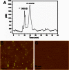
Isolation and characterization of Tau oligomers from TBI brains. Tau oligomers were isolated by immunoprecipitation (IP) using anti-Tau oligomer antibody T22 from rat brains extracted 24 h after TBI. A, size exclusion chromatogram of Tau oligomers showing two main peaks, one at 75–150 kDa, which may include Tau dimer/trimers, and a second peak at 300 kDa. B, AFM image of the TBI brain-derived Tau oligomers. C, AFM image of the material isolated from sham brains.
FIGURE 3.
FIGURE 3.
Tau oligomer accumulation in hippocampus and cortex in TBI. A–C, representative immunofluorescent images of hippocampus 24 h post-TBI showing Tau-1 (A, green), T22 (B, red), and merged signals (C, also including DAPI (blue)), confirming the presence of Tau oligomers in situ. D–F, representative immunofluorescent images of sham (24 h) hippocampal sections. G–I, representative immunofluorescent photomicrographs of cortex sections post TBI (24 h) showing Tau 1 (G, green) and T22 (H, red arrows indicate representative positive pyramidal neurons), and merged images (I, also including DAPI (blue)). J–L, representative immunofluorescent images of sham (24 h) cortical sections. Scale bar, 25 μm.
FIGURE 4.
FIGURE 4.
Detection of Tau oligomers within 24 h of TBI. A–F, phosphorylated Tau (AT180) and Tau oligomers (T22) were detected in TBI brains (24-h survival) using peroxidase immunohistochemistry in the CA3 (A and D), dentate gyrus (B and E), and cortex (C and F). Scale bar, 10 μm.
FIGURE 5.
FIGURE 5.
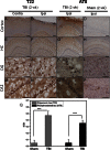
Detection of Tau oligomers 2 weeks after TBI. Tau oligomers (T22) and phosphorylated Tau (AT8) were detected in TBI brains (2-week (2 wk) survival) using peroxidase immunohistochemistry in the cortex (contralateral (Contra) and ipsilateral (Ipsi) to the injury site) and in the dentate gyrus (DG) and CA1/2 (CA2) regions of the hippocampus (HC). Control sham-injured brains were stained with AT8. Scale bars, 200 μm (A–H) and 50 μm (I–P). Q, Tau oligomers and phosphorylated Tau were quantified by direct ELISA (PBS extracts) using T22 and AT8, respectively. Samples measured in triplicates (n = 2); ***, p < 0.0005.
FIGURE 6.
FIGURE 6.
Schematic showing a proposed mechanism regarding the means by which neuronal injuries and cell death resulting from TBI may lead to rapid formation of toxic Tau oligomers, which may initiate the spread of Tau pathology.

Similar articles

Cited by

References

    1. Colicos M. A., Dixon C. E., Dash P. K. (1996) Delayed, selective neuronal death following experimental cortical impact injury in rats: possible role in memory deficits. Brain Res. 739, 111–119 - PubMed
    1. Smith D. H., Chen X. H., Pierce J. E., Wolf J. A., Trojanowski J. Q., Graham D. I., McIntosh T. K. (1997) Progressive atrophy and neuron death for one year following brain trauma in the rat. J. Neurotrauma 14, 715–727 - PubMed
    1. McKinney R. A., Debanne D., Gähwiler B. H., Thompson S. M. (1997) Lesion-induced axonal sprouting and hyperexcitability in the hippocampus in vitro: implications for the genesis of posttraumatic epilepsy. Nat. Med. 3, 990–996 - PubMed
    1. Pitkänen A., McIntosh T. K. (2006) Animal models of post-traumatic epilepsy. J. Neurotrauma 23, 241–261 - PubMed
    1. Behan L. A., Phillips J., Thompson C. J., Agha A. (2008) Neuroendocrine disorders after traumatic brain injury. J. Neurol. Neurosurg. Psychiatry 79, 753–759 - PubMed

Publication types

LinkOut - more resources