

Review
doi: 10.1016/j.sbi.2013.04.002.
Epub 2013 Apr 27.
Structural studies on the regulation of Ca2+/calmodulin dependent protein kinase II
Affiliations
- PMID: 23632248
- PMCID: PMC3788167
- DOI: 10.1016/j.sbi.2013.04.002
Item in Clipboard
Review
Structural studies on the regulation of Ca2+/calmodulin dependent protein kinase II
Curr Opin Struct Biol.
2013 Apr.
Abstract
Ca(2+)/calmodulin dependent protein kinase II (CaMKII) is a broadly distributed metazoan Ser/Thr protein kinase that is important in neuronal and cardiac signaling. CaMKII forms oligomeric assemblies, typically dodecameric, in which the calcium-responsive kinase domains are organized around a central hub. We review the results of crystallographic analyses of CaMKII, including the recently determined structure of a full-length and autoinhibited form of the holoenzyme. These structures, when combined with other data, allow informed speculation about how CaMKII escapes calcium-dependence when calcium spikes exceed threshold frequencies.
Copyright © 2013 Elsevier Ltd. All rights reserved.
Figures
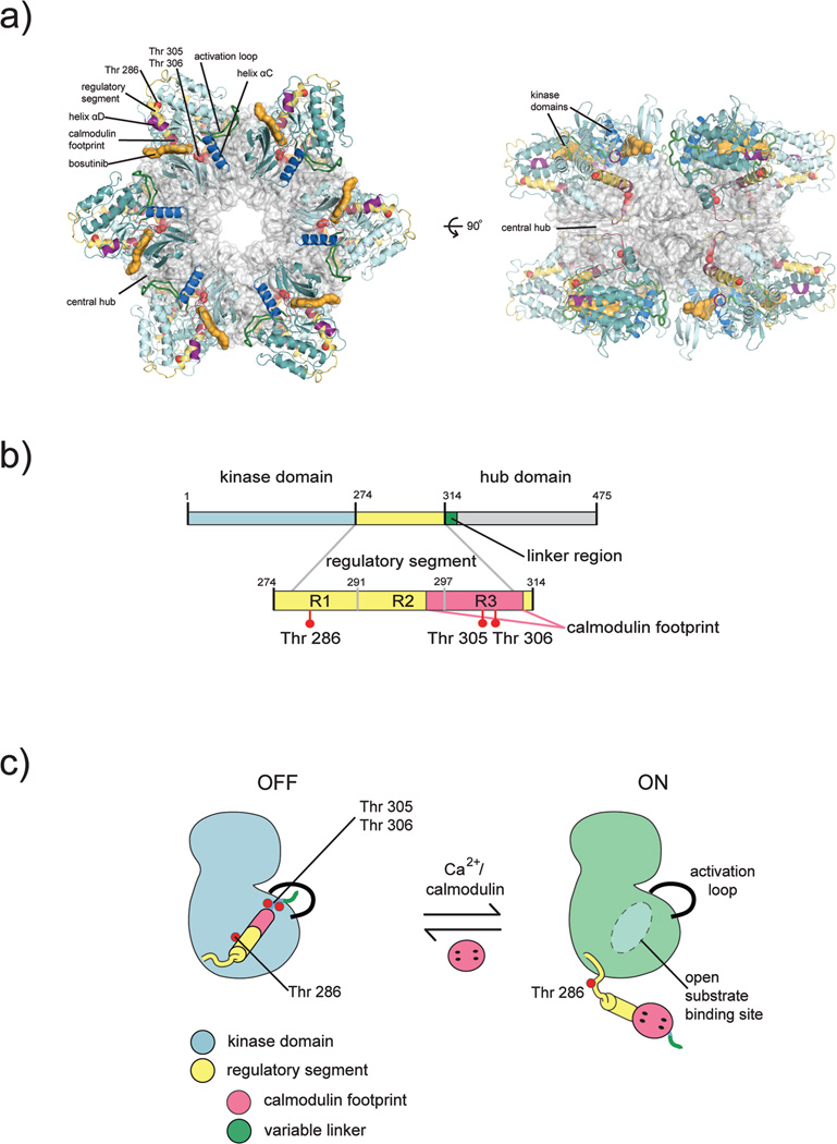
Structural organization of CaMKII. a) Crystal structure of an intact CaMKII holoenzyme. The hub assembly is shown as a surface representation (gray) and the kinase domains are shown as cartoons. Each kinase domain is nestled between two hub domains, making contacts with each. The CaM binding region is buried by interactions with the hub. b) Each CaMKII subunit is comprised of a kinase domain (blue), regulatory segment (yellow), variable linker region (green), and a hub domain (gray). The regulatory segment contains three critical phosphorylation sites: Thr 286 is located within the R1 region of the regulatory segment, and Thr 305, Thr 306 are located within the CaM binding region (pink). The CaM binding footprint spans part of the R2 region (docking site for the catalytic domain) and the entire R3 region within the regulatory segment. c) Activation by Ca2+/CaM displaces the regulatory segment from its docking site, thereby freeing the substrate binding site and exposing Thr 286 for trans phosphorylation by an adjacent CaMKII kinase. This figure was adapted from (35).
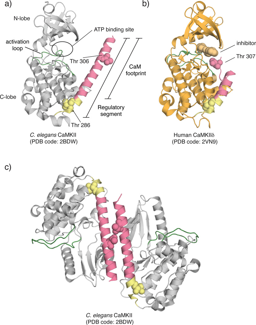
Crystal structures of kinase domains of CaMKII variants. a) C. elegans CaMKII. The regulatory segment is completely helical and projects Thr 306 far from the active site. PDB code: 2BDW b) Human CaMKIIδ crystallized bound to a small molecule inhibitor, Bisindolylmaleimide IX. The residues of the CaM binding region form a flexible loop instead of a helix as seen in (a). Colors correspond to those in (a). PDB code: 2V7O c) Crystallized dimer of C. elegans CaMKII. The regulatory segments of two kinases form a coiled-coil, burying the CaM binding region.
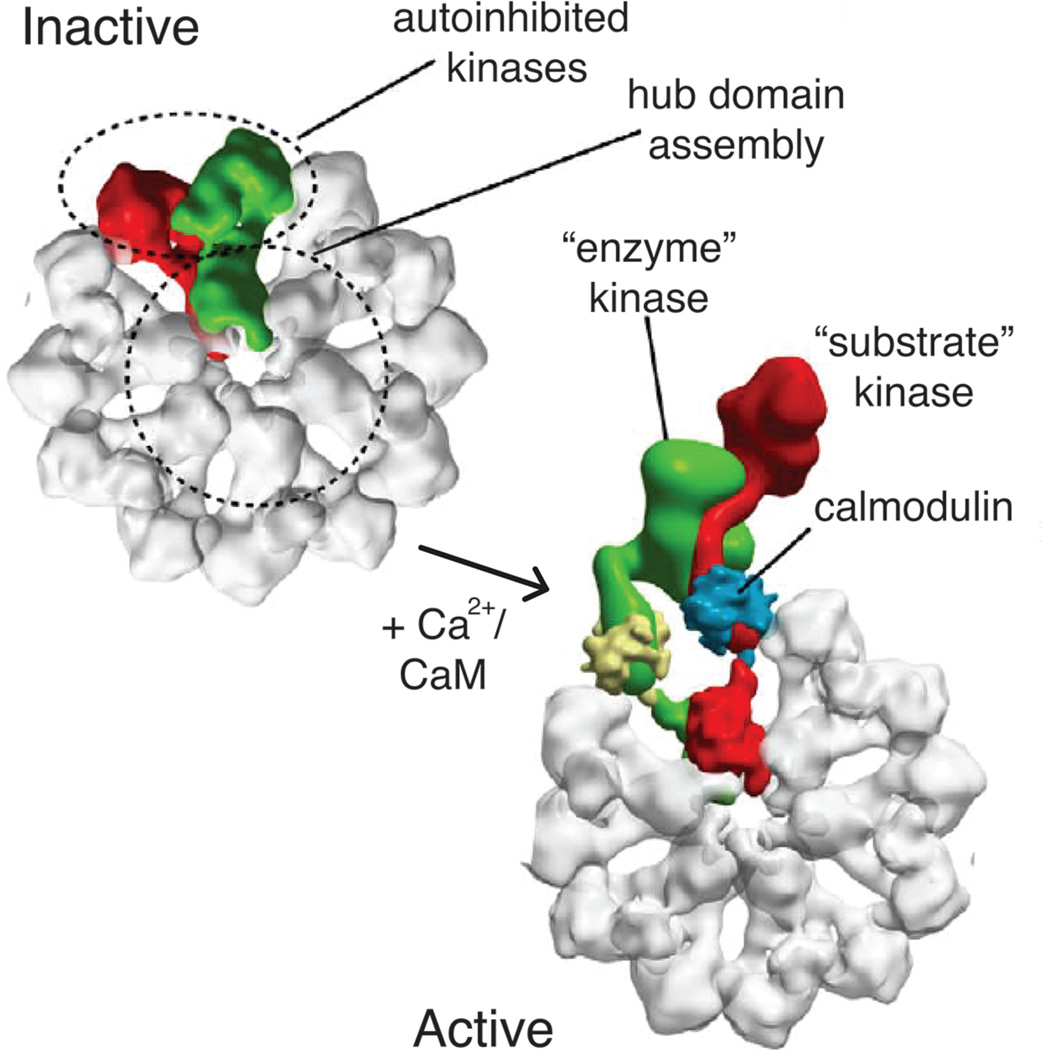
Substrate capture during activation within the CaMKII holoenzyme. In the inactive state, each kinase is autoinhibited. Upon Ca2+/CaM binding, activation occurs cooperatively within the holoenzyme via a substrate capture mechanism. An enzyme kinase captures a substrate kinase (both bound to CaM) and results in Thr 286 trans autophosphorylation. This figure was adapted from (39) with permissions.
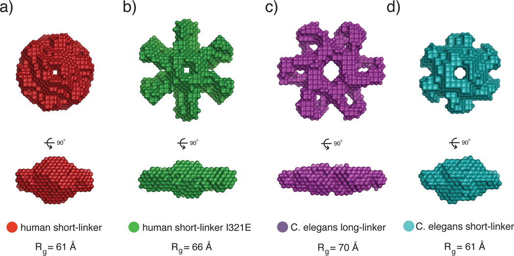
SAXS analysis of the CaMKII holoenzyme. a) The human short-linker construct, used for the crystallization of the full-length holoenzyme, has a compact SAXS envelope. b) A mutation (I321E) introduced to the short-linker construct at the docking site between the kinase and hub domain shows a significantly larger SAXS envelope. This may represent an extended form of the autoinhibited holoenzyme where the kinases are popped out from the hub domain. Additionally, the I321E has a left-shifted EC50 value for CaM binding, which corroborates the model that the extended conformation is more accessible to CaM than the compact conformation observed in the crystal structure. c,d) C. elegans CaMKII also converts from a large to small SAXS envelope when the variable linker is shortened. These may also represent the extended and compact conformations of CaMKII, respectively. This figure was adapted from (35).
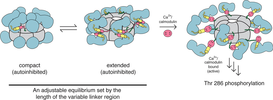
Conformational changes in the holoenzyme. The CaMKII holoenzyme has been shown to adopt both compact (crystallization, SAXS) and extended (SAXS) autoinhibited states. An equilibrium exists between these states, and is sensitive to the length of the variable linker connecting the kinase and hub domains. In the compact autoinhibited conformation, residues comprising the CaM binding region make contacts with the hub domain, thereby making it highly inaccessible to CaM. In the extended autoinhibited conformation, the CaM binding region is more exposed, though the structural details of this extended state have yet to be elucidated. Upon Ca2+/CaM binding, the regulatory segment is released from the kinase domain, thereby relieving inhibition. Active kinases then trans autophosphorylate adjacent kinases and subsequently phosphorylate downstream targets. Colors correspond to those in Fig. 1. This figure was adapted from (35).
References
-
- Kemp BE, Parker MW, Hu S, Tiganis T, House C. Substrate and pseudosubstrate interactions with protein kinases: determinants of specificity. Trends Biochem Sci. 1994;19:440–444. - PubMed
-
- Heierhorst J, Kobe B, Feil SC, Parker MW, Benian GM, Weiss KR, Kemp BE. Ca2+/S100 regulation of giant protein kinases. Nature. 1996;380:636–639. - PubMed
-
- Owen DJ, Noble ME, Garman EF, Papageorgiou AC, Johnson LN. Two structures of the catalytic domain of phosphorylase kinase: an active protein kinase complexed with substrate analogue and product. Structure. 1995;3:467–482. - PubMed
-
- Goldberg J, Nairn AC, Kuriyan J. Structural basis for the autoinhibition of calcium/calmodulin-dependent protein kinase I. Cell. 1996;84:875–887. - PubMed
Publication types
MeSH terms
Substances
Grants and funding
LinkOut - more resources
Full Text Sources
Other Literature Sources
Molecular Biology Databases
Miscellaneous
