Review
. 2014 Jan;42(1):16-24.
doi: 10.1016/j.dci.2013.04.011. Epub 2013 Apr 28.

Conventional and non-conventional Drosophila Toll signaling

Affiliations
Review

Conventional and non-conventional Drosophila Toll signaling

Scott A Lindsay et al. Dev Comp Immunol. 2014 Jan.

Abstract

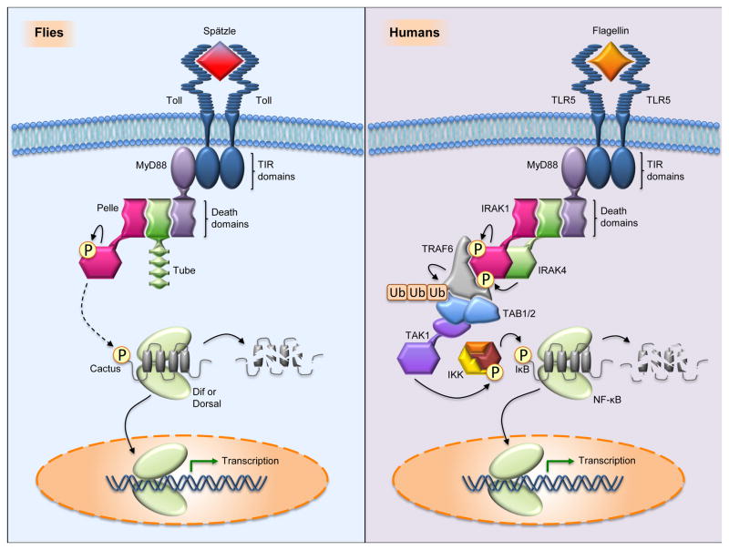
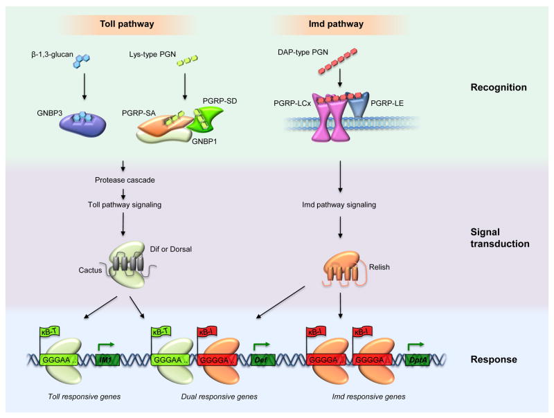
The discovery of Toll in Drosophila and of the remarkable conservation in pathway composition and organization catalyzed a transformation in our understanding of innate immune recognition and response. At the center of that picture is a cascade of interactions in which specific microbial cues activate Toll receptors, which then transmit signals driving transcription factor nuclear localization and activity. Experiments gave substance to the vision of pattern recognition receptors, linked phenomena in development, gene regulation, and immunity into a coherent whole, and revealed a rich set of variations for identifying non-self and responding effectively. More recently, research in Drosophila has illuminated the positive and negative regulation of Toll activation, the organization of signaling events at and beneath membranes, the sorting of information flow, and the existence of non-conventional signaling via Toll-related receptors. Here, we provide an overview of the Toll pathway of flies and highlight these ongoing realms of research.

Keywords: Drosophila; Innate immunity; NF-κB; Non-conventional pathway; Toll.


Figures

Fig. 1
Fig. 1
Evolutionary conservation of the Drosophila Toll and human TLR signaling pathways. (left) In flies, Toll signaling is activated when a processed form of Spätzle binds the Toll ectodomain. Toll activation triggers dimerization of the intracytoplasmic TIR domains, which promotes binding of the adaptor protein MyD88 through its own TIR domain. MyD88 binds the adaptor protein Tube, which in turn recruits the protein kinase Pelle, each interaction occurring via pairwise interaction of death domains. Although only one signaling module is shown, each TIR domain of the Toll dimer is capable of binding one molecule of MyD88 and there are thus two signaling modules per Toll dimer. Recruitment of Pelle induces its autophosphorylation, triggering phosphorylation and destruction of the inhibitor Cactus. The transcription factor, either Dif or Dorsal depending on the context, is then freed for nuclear translocation. (right) In humans, there are numerous TLR pathways involving often distinct but sometimes overlapping sets of PAMPs, signaling components, and transcription factors. In the example illustrated, TLR5 signaling is activated by Flagellin, a principal component of bacterial flagella. In a manner analogous to Drosophila Toll signaling, human MyD88 builds a signaling complex with the Tube ortholog, IRAK4, and the Pelle ortholog, IRAK1. The complex is much bigger than the Drosophila counterpart, comprising 6 MyD88, 4 IRAK4, and 4 IRAK1 molecules in a complete signaling unit. IRAK4 phosphorylates IRAK1, triggering IRAK1 autophosphorylation and dissociation from the complex. Activated IRAK1 binds TRAF6, which then autoubiquitinates and binds the TAB/TAK1 proteins. TAK1 becomes activated and phosphorylates the IKK complex, which then phosphorylates the inhibitor IκB, leading to its degradation and the nuclear translocation of NF-κB.
Fig. 2
Fig. 2
Protease cascades leading to Toll activation. The active form of the Toll ligand Spätzle results from a specific cleavage triggered by any of four serine protease cascades. In these illustrations, horizontal red arrows denote proteolytic conversion of the zymogens to their active forms and a reddish glow denotes the active form of a protease. (left) In early embryogenesis, positional cues laid out during oogenesis establish the dorsoventral axis through the localized activation of Toll on the ventral side of the embryo. The protease cascade that triggers this Toll activation involves Nudel, Gastrulation defective (gd), Snake, and Easter. Nudel, directly or indirectly activates the Gastrulation defective protease, which then activates Snake. With the involvement of the sulfotransferase Pipe, activated Snake cleaves and activates Easter. Activated Easter processes Spätzle, completing generation of a functional ligand for Toll. (middle, right) A similar mechanism operates in innate immunity, where three protease cascades converge at the activation step for the Spätzle processing enzyme (SPE). In the case of fungi and Gram-positive bacteria, the cell wall components β-1,3-glucan and Lys-type peptidoglycan, respectively, are recognized by circulating pathogen recognition receptors and trigger separate, but related protease cascades. The serine protease ModSP integrates signals from these recognition molecules and activates the protease Grass, which activates SPE. Other immune factors, such as the serine proteases Spirit, Sphinx, and Spheroide may function between Grass and SPE. In addition to recognizing PAMPs, the innate immune system is capable of sensing fungi and bacteria via the zymogen Persephone. Virulence factors (proteases) secreted from microbes cleave Persephone, resulting in activation of SPE. At several points in the pathways shown, serpins are known to provide negative regulation of these immune protease cascades. Necrotic inhibits Persephone and Spn1 inhibits upstream of Grass, with ModSP a likely target.
Fig. 3
Fig. 3
Specificity and synergy in Toll and Imd signaling. The Toll and Imd pathways recognize distinct PAMPs and generate distinct responses, with either separate or coordinate regulation of transcriptional outputs. In the case of fungi and most Gram-positive bacteria, cell wall components (β-1,3,-glucan from fungi; Lys-type peptidoglycan from bacteria) are recognized by extracellular pathogen recognition receptors and the signal is transduced through protease cascades to activate Toll pathway signaling. A pure Toll response involves binding of a homodimer of the transcription factor Dif or Dorsal, typically at a single Toll-specific κB site (κB-T) upstream of Toll-responsive genes. In the case of Gram-negative bacteria, and select Gram-positive species, polymeric DAP-type peptidoglycan (PGN) is recognized by a dimer of PGRP-LCx and an extracellular version of PGRP-LE (containing only the PGRP domain) to activate Imd signaling. A pure Imd response involves two or more homodimers of the transcription factor Relish binding at neighboring Imd-specific κB sites (κB-I) upstream of Imd-responsive genes. In the event that both pathways are stimulated, Toll- and Imd-regulated Rel proteins can cooperatively regulate a third set of genes. The promoters of such dual-responsive genes contain neighboring Toll-specific and Imd-specific κB sites, where homodimers of Dif or Dorsal and of Relish, respectively, can bind to effect transcription. IM1: Immune-induced molecule 1. Def: Defensin. DptA: Diptericin A.

References

    1. Akhouayri I, Turc C, Royet J, Charroux B. Toll-8/Tollo negatively regulates antimicrobial response in the Drosophila respiratory epithelium. PLoS Pathog. 2011;7:e1002319. - PMC - PubMed
    1. Anderson KV, Nusslein VC. Information for the dorsal-ventral pattern of the Drosophila embryo is stored as maternal mRNA. Nature. 1984;311:223–227. - PubMed
    1. Ayyar S, Pistillo D, Calleja M, Brookfield A, Gittins K, Goldstone C, Simpson P. NF-kappaB/Rel-mediated regulation of the neural fate in Drosophila. PLoS One. 2007;2:e1178. - PMC - PubMed
    1. Belvin MP, Anderson KV. A conserved signaling pathway: the Drosophila toll-dorsal pathway. Ann Rev Cell Dev Biol. 1996;12:393–416. - PubMed
    1. Bilak H, Tauszig-Delamasure S, Imler JL. Toll and Toll-like receptors in Drosophila. Biochem Soc Trans. 2003;31:648–651. - PubMed

Publication types