. 2013 Jun;131(6):1644-52.
doi: 10.1016/j.jaci.2013.03.002. Epub 2013 Apr 28.

Compartmentalized chemokine-dependent regulatory T-cell inhibition of allergic pulmonary inflammation

Affiliations

Compartmentalized chemokine-dependent regulatory T-cell inhibition of allergic pulmonary inflammation

Roshi Afshar et al. J Allergy Clin Immunol. 2013 Jun.

Abstract

Background: Induction of endogenous regulatory T (Treg) cells represents an exciting new potential modality for treating allergic diseases, such as asthma. Treg cells have been implicated in the regulation of asthma, but the anatomic location in which they exert their regulatory function and the mechanisms controlling the migration necessary for their suppressive function in asthma are not known. Understanding these aspects of Treg cell biology will be important for harnessing their power in the clinic.

Objective: We sought to determine the anatomic location at which Treg cells exert their regulatory function in the sensitization and effector phases of allergic asthma and to determine the chemokine receptors that control the migration of Treg cells to these sites in vivo in both mice and human subjects.

Methods: The clinical efficacy and anatomic location of adoptively transferred chemokine receptor-deficient CD4(+)CD25(+) forkhead box protein 3-positive Treg cells was determined in the sensitization and effector phases of allergic airway inflammation in mice. The chemokine receptor expression profile was determined on Treg cells recruited into the human airway after bronchoscopic segmental allergen challenge of asthmatic patients.

Results: We show that CCR7, but not CCR4, is required on Treg cells to suppress allergic airway inflammation during the sensitization phase. In contrast, CCR4, but not CCR7, is required on Treg cells to suppress allergic airway inflammation during the effector phase. Consistent with our murine studies, human subjects with allergic asthma had an increase in CCR4-expressing functional Treg cells in the lungs after segmental allergen challenge.

Conclusion: The location of Treg cell function differs during allergic sensitization and allergen-induced recall responses in the lung, and this differential localization is critically dependent on differential chemokine function.


Figures

Figure 1
Figure 1. CCR7, but not CCR4, is required on Tregs to suppress allergic airway inflammation in the sensitization model
(a) Schematic of sensitization model of Treg suppression (see Methods for details). (b) Total cells, eosinophils and lymphocytes in the BAL were enumerated at day 21 in WT mice that received PBS (Solid bars), WT OT-II Tregs (grey bars), CCR4−/− OT-II Tregs (checked bars), or CCR7−/− OT-II Tregs (white bars) i.v. on day -1 followed by OVA i.p on days 0 and 14. Data are representative of three experiments. (c) Histopathological analysis of lung sections on day 21 stained with H&E (top) and PAS (bottom). Scale bars, 50μm. Data are representative of 3 independent experiments. (d) Blinded histolopathological scoring of lung inflammation on day 21 from mice treated as above (*p<0.05 compared to PBS injected mice; n ≥ 10 mice per group from three experiments). (e) Cytokines measured in the BAL. Data are mean ± SEM of 8 mice per group from two experiments (*p<0.05).
Figure 2
Figure 2. CCR4, but not CCR7, is required on Tregs to suppress allergic airway inflammation in the effector model
(a) Schematic of the effector model of Treg suppression (see Methods for details). (b) Total cells, eosinophils and lymphocytes in the BAL were enumerated on day 21 in OVA immunized mice that received PBS (Solid bars), WT OT-II Tregs (grey bars), CCR4−/− OT-II Tregs (checked bars), or CCR7−/− OT-II Tregs (white bars) on day 16. Data are representative of 3 experiments. (c) Histopathological analysis of lung sections stained with H&E (top) and PAS (bottom). Scale of bar 50μm. Data are representative of 3 independent experiments. (d) Blinded histopathological scoring of lung inflammation on day 21 from mice treated as above (*p<0.05 compared to PBS injected mice; n ≥10 mice per group from 3 experiments). (e) Cytokines measured in the BAL. Data are a mean ± SEM of 8 mice per group from 2 experiments (*p<0.05).
Figure 3
Figure 3. Chemokine receptor-dependent trafficking of Tregs to LN and lung
(a) Schematic of experimental protocol. (b) Cells from thoracic LN and lung were analyzed for Foxp3+Thy1.2+ expression (gated on CD4+CD25+ cells) on day 5. Data are pooled from 4 independent experiments. (c) Chemokine expression in LNs (pooled thoracic and inguinal) and lungs from WT mice measured on day 5 by RT-QPCR (n=8 mice from 2 independent experiments) (d) Schematic of experimental protocol. (e) Percent of Foxp3+Thy1.2+ cells of CD4+CD25+Foxp3+ cells in lymphocyte gate isolated from the LN and lung on day 19. These data are pooled from four independent experiments. (f) Chemokine expression in the thoracic LN and lung from WT mice measured by RT-QPCR on day 19 (n=8 mice from 2 independent experiments). (g) Percent CCR4 and CCR7 cell surface expression on Tregs (gated on CD4+CD25+Foxp3+ cells in lymphocyte gate) recovered from the lung on day 21 in the effector model. Data are mean ± SEM.
Figure 4
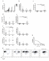
Figure 4. Tregs recruited into the human allergic lung express CCR4 and are functional
Data from 10 allergic asthmatic subjects (a-f). (a) Number of lymphocytes, monocytes, neutrophils, and eosinophils from baseline and allergen challenge BAL (mean ± SEM ). (b) The percent and (c) number of CD4+ cells recovered from the BAL pre-allergen challenge (pre) and 24 hours post-bronchoscopic segmental diluent (dil) or allergen (Ag) challenge (*p<0.05; n=10 subjects). (d) The percent of CD25+Foxp3+ Tregs of total CD4+ cells and (e) the number of CD4+CD25+Foxp3+ Tregs recovered from BAL pre- and 24 hours post bronchoscopic diluent or allergen challenge (*p<0.05; n=10 subjects). (f) Percent of Tregs (CD4+CD25+Foxp3+) expressing CCR4, CCR6, CCR7 and CXCR3 in the BAL. (*p<0.05; n=10 subjects). (g, h) Treg suppression assay. (g) Representative FACS plots and (h) quantitation of percent inhibition of T effector (Teff) proliferation in the presence or varying ratios of Teff to Tregs (n=3).

References

    1. Locksley RM. Asthma and allergic inflammation. Cell. 2010;140:777–83. - PMC - PubMed
    1. Larche M, Robinson DS, Kay AB. The role of T lymphocytes in the pathogenesis of asthma. J Allergy Clin Immunol. 2003;111:450–63. quiz 64. - PubMed
    1. Lahl K, Loddenkemper C, Drouin C, Freyer J, Arnason J, Eberl G, et al. Selective depletion of Foxp3+ regulatory T cells induces a scurfy-like disease. J Exp Med. 2007;204:57–63. - PMC - PubMed
    1. Strickland DH, Stumbles PA, Zosky GR, Subrata LS, Thomas JA, Turner DJ, et al. Reversal of airway hyperresponsiveness by induction of airway mucosal CD4+CD25+ regulatory T cells. J Exp Med. 2006;203:2649–60. - PMC - PubMed
    1. Kearley J, Barker JE, Robinson DS, Lloyd CM. Resolution of airway inflammation and hyperreactivity after in vivo transfer of CD4+CD25+ regulatory T cells is interleukin 10 dependent. J Exp Med. 2005;202:1539–47. - PMC - PubMed

Publication types

MeSH terms