Review
. 2013 Sep;61(9):1379-401.
doi: 10.1002/glia.22500. Epub 2013 Apr 30.

How stem cells speak with host immune cells in inflammatory brain diseases

Affiliations
Review

How stem cells speak with host immune cells in inflammatory brain diseases

Stefano Pluchino et al. Glia. 2013 Sep.

Abstract

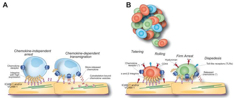
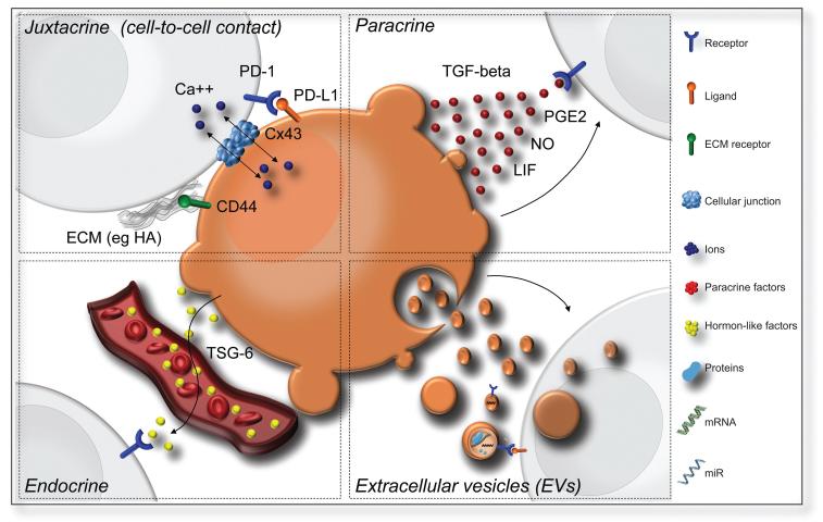
Advances in stem cell biology have raised great expectations that diseases and injuries of the central nervous system (CNS) may be ameliorated by the development of non-hematopoietic stem cell medicines. Yet, the application of adult stem cells as CNS therapeutics is challenging and the interpretation of some of the outcomes ambiguous. In fact, the initial idea that stem cell transplants work only via structural cell replacement has been challenged by the observation of consistent cellular signaling between the graft and the host. Cellular signaling is the foundation of coordinated actions and flexible responses, and arises via networks of exchanging and interacting molecules that transmit patterns of information between cells. Sustained stem cell graft-to-host communication leads to remarkable trophic effects on endogenous brain cells and beneficial modulatory actions on innate and adaptive immune responses in vivo, ultimately promoting the healing of the injured CNS. Among a number of adult stem cell types, mesenchymal stem cells (MSCs) and neural stem/precursor cells (NPCs) are being extensively investigated for their ability to signal to the immune system upon transplantation in experimental CNS diseases. Here, we focus on the main cellular signaling pathways that grafted MSCs and NPCs use to establish a therapeutically relevant cross talk with host immune cells, while examining the role of inflammation in regulating some of the bidirectionality of these communications. We propose that the identification of the players involved in stem cell signaling might contribute to the development of innovative, high clinical impact therapeutics for inflammatory CNS diseases.

Keywords: immune modulation; inflammation; mesenchymal stem cells; neural stem cells; stem cell-host interactions.


Figures

FIGURE 1
FIGURE 1
Rules for the migration of effector T cells and systemically injected stem cells to the CNS. (A) Activated effector T lymphocytes have a high expression of membrane-bound integrins that are able to support chemokine-independent arrest under flow. The transmigration of effector T cells requires chemokines that are stored in intracellular vesicles, ready to be released in close contact with crawling cells. (B) Tethering, rolling, and firm arrest of injected stem cells to activated endothelial cells and diapedesis into inflamed CNS areas are sequentially mediated by the constitutive expression of functional α and β integrins, cell adhesion molecules such as CD44, TLRs and chemokine receptors on the MSC/NPC surface. *The main chemokine signaling pathways responsible for stem cell migration are shown in Table 2.
FIGURE 2
FIGURE 2
NPCs express environmental sensors. (A) Z-stack confocal image (from a total of n5 40 Z-stacks of optical slices in 0.5 μm intervals) of a mouse neurosphere in vitro. Red is for vimentin, green is for CD44, white is for phosphor-histone H3 (pHH3) and blue is for cell nuclei (Dapi). (B) Volocity V®-based 3D reconstruction of the CD44 expression in A. The magnified frame in A shows a pHH3+/CD44+ NPC.
FIGURE 3
FIGURE 3
Schematic representation of the alternative cell signaling pathways regulating the interactions between the stem cell graft and the host immune system in inflammatory brain diseases. (A) Juxtacrine signaling pathways (cell-to-cell contact) that include (i) receptor-ligand interaction such as PD-L1/PD-1; (ii) receptor binding to components of the extracellular matrix (ECM) released by neighboring cells (such as CD44 to HA); and iii) gap junction formation (via connexins); (B) Paracrine signaling with release of soluble factors that likely form a gradient (e.g. TGF-β, LIF, NO, PGE2); (C) Endocrine signaling (signaling at a distance) that implies the release of hormonelike factors such as TSG-6; and (D) EV release with the possibility to deliver a multitude of bioactive molecules such as mRNAs, micro-RNAs and proteins. Abbreviations: CD; cluster of differentiation; HA: hyaluronic acid; ECM: extracellular matrix; Cx43: Connexin-43; Ca++: calcium; TGF-β: transforming growth factor beta; PGE2: Prostaglandin E2; NO: nitric oxide; LIF: Leukaemia inhibitory factor; TSG-6: TNF-α-stimulated gene/protein 6; PD-1: programmed death-1; PD-L1: programmed cell death 1 ligand 1; EVs: extracellular vesicles.
FIGURE 4
FIGURE 4
NPC grafts establish juxtacrine signaling with endogenous professional phagocytes through junctional coupling. (A) Confocal microscopy image of GFP (green) NPCs contacting F4/80+ macrophages via connexin43+ cellular junctions (red; arrowheads). (B) Volocity V®-based 3D reconstruction of the confocal Z-stack in A. The magnified inset shows a structural junctional connexin43 pattern (red; arrowheads) between the process of one NPC (green) and one juxtaposed F4/80+ macrophage (blue). (C) Immunoelectron micrograph of GFP+ NPCs. The frame indicates one NPC whose processes are found to be in very close contact with a (GFP) monocyte/macrophage. (D) High magnification of the frame in C showing the immunogold-labeled process of an NPC (arrowheads) running between a monocyte/macrophage and a second immunogold-labeled NPC. Cellular junctions between both NPC cytoplasms (inset, arrows) and between the NPC and the monocyte/macrophage can be observed in the inset. Pseudo colors in B and C: NPCs are in green; monocytes/macrophages are in orange; endothelial cells are in yellow and endogenous astrocytes are in blue. (Reproduced with permission from Cusimano et al., Brain, 2012, 135, 447-460, ©Oxford University Press.)

References

    1. Aboody K, Capela A, Niazi N, Stern JH, Temple S. Translating stem cell studies to the clinic for CNS repair: Current state of the art and the need for a Rosetta Stone. Neuron. 2011;70:597–613. - PubMed
    1. Aboody KS, Brown A, Rainov NG, Bower KA, Liu S, Yang W, Small JE, Herrlinger U, Ourednik V, Black PM, Breakefield XO, Snyder EY. Neural stem cells display extensive tropism for pathology in adult brain: evidence from intracranial gliomas. Proc Natl Acad Sci USA. 2000;97:12846–12851. - PMC - PubMed
    1. Aggarwal S, Pittenger MF. Human mesenchymal stem cells modulate allogeneic immune cell responses. Blood. 2005;105:1815–1822. - PubMed
    1. Ahmed AU, Thaci B, Alexiades NG, Han Y, Qian S, Liu F, Balyasnikova IV, Ulasov IY, Aboody KS, Lesniak MS. Neural stem cell-based cell carriers enhance therapeutic efficacy of an oncolytic adenovirus in an orthotopic mouse model of human glioblastoma. Mol Ther. 2011;19:1714–1726. - PMC - PubMed
    1. Akiyama K, Chen C, Wang D, Xu X, Qu C, Yamaza T, Cai T, Chen W, Sun L, Shi S. Mesenchymal-stem-cell-induced immunoregulation involves FAS-ligand-/FAS-mediated T cell apoptosis. Cell Stem Cell. 2012;10:544–555. - PMC - PubMed

Publication types