. 2013 Jun 15;73(12):3591-603.
doi: 10.1158/0008-5472.CAN-12-4100. Epub 2013 Apr 30.

Dual blockade of PD-1 and CTLA-4 combined with tumor vaccine effectively restores T-cell rejection function in tumors

Affiliations

Dual blockade of PD-1 and CTLA-4 combined with tumor vaccine effectively restores T-cell rejection function in tumors

Jaikumar Duraiswamy et al. Cancer Res. .

Abstract

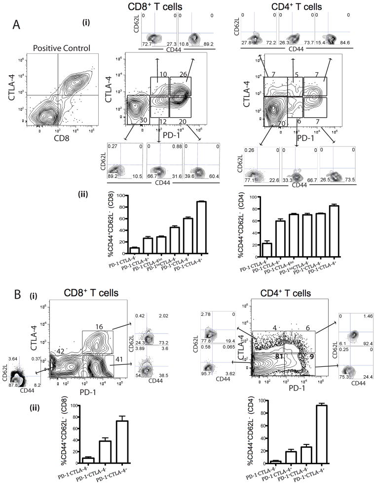
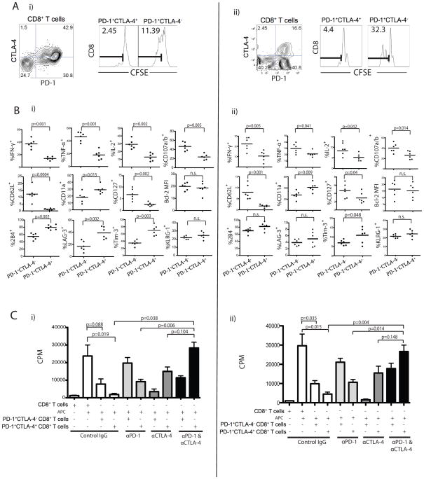
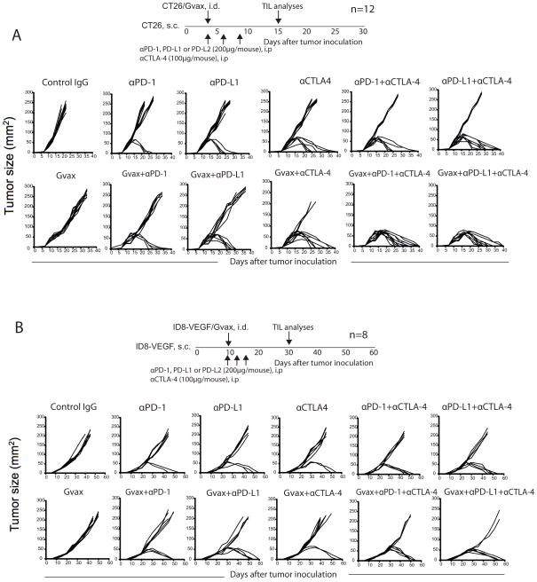
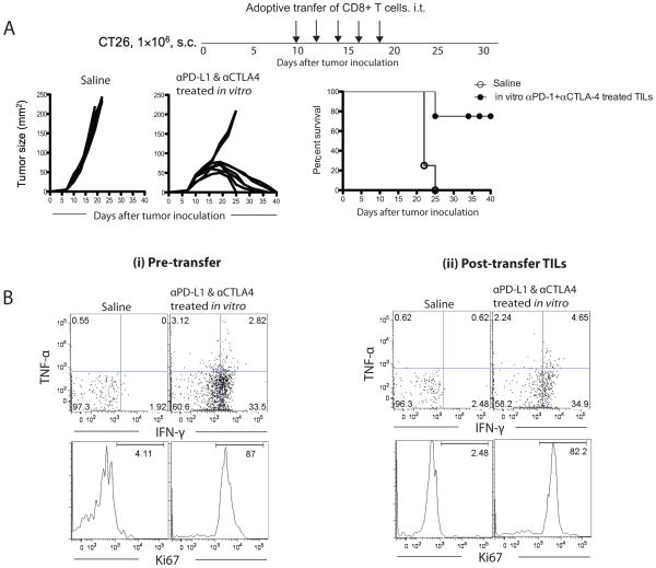
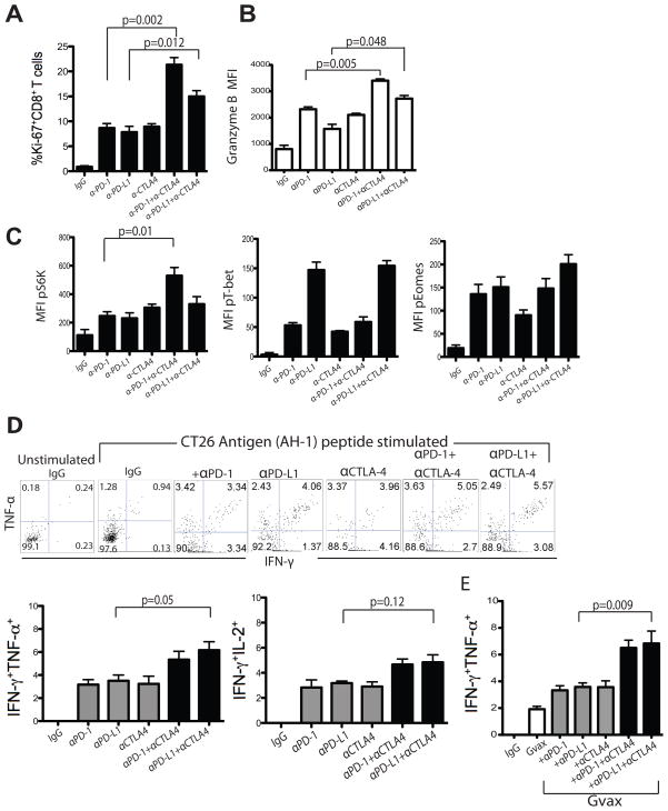
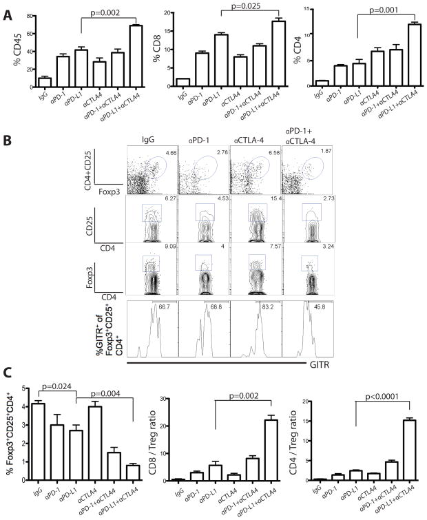
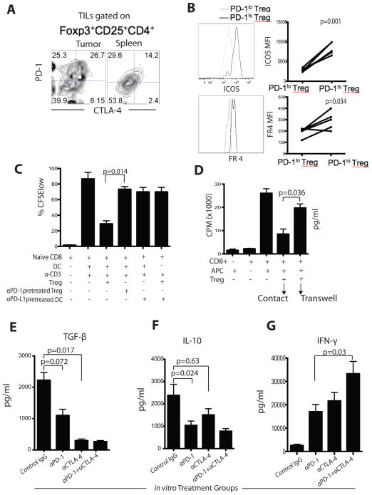
Tumor progression is facilitated by regulatory T cells (Treg) and restricted by effector T cells. In this study, we document parallel regulation of CD8(+) T cells and Foxp3(+) Tregs by programmed death-1 (PD-1, PDCD1). In addition, we identify an additional role of CTL antigen-4 (CTLA-4) inhibitory receptor in further promoting dysfunction of CD8(+) T effector cells in tumor models (CT26 colon carcinoma and ID8-VEGF ovarian carcinoma). Two thirds of CD8(+) tumor-infiltrating lymphocytes (TIL) expressed PD-1, whereas one third to half of CD8(+) TIL coexpressed PD-1 and CTLA-4. Double-positive (PD-1(+)CTLA-4(+)) CD8(+) TIL had characteristics of more severe dysfunction than single-positive (PD-1(+) or CTLA-4(+)) TIL, including an inability to proliferate and secrete effector cytokines. Blockade of both PD-1 and CTLA-4 resulted in reversal of CD8(+) TIL dysfunction and led to tumor rejection in two thirds of mice. Double blockade was associated with increased proliferation of antigen-specific effector CD8(+) and CD4(+) T cells, antigen-specific cytokine release, inhibition of suppressive functions of Tregs, and upregulation of key signaling molecules critical for T-cell function. When used in combination with GVAX vaccination (consisting of granulocyte macrophage colony-stimulating factor-expressing irradiated tumor cells), inhibitory pathway blockade induced rejection of CT26 tumors in 100% of mice and ID8-VEGF tumors in 75% of mice. Our study indicates that PD-1 signaling in tumors is required for both suppressing effector T cells and maintaining tumor Tregs, and that PD-1/PD-L1 pathway (CD274) blockade augments tumor inhibition by increasing effector T-cell activity, thereby attenuating Treg suppression.


Figures

Figure 1
Figure 1. Phenotypic analysis of CT26 and ID8-VEGF TIL
TIL isolated from CT26 and ID8-VEGF tumors were stained with antibodies to CD45, CD8, CD4, PD-1, CTLA-4, CD44, and CD62L as well as live/dead stain. Representative CT26 (A) and ID8-VEGF (B) tumor samples showing expression of PD-1 and CTLA-4 on CD8+CD45+ and CD4+CD45+ TIL with distinct cell populations, which are further analyzed for CD44 and CD62L. Summary data of CTLA-4 and PD-1 expression on TEM (CD44hiCD62Llo) CD8+ and CD4+ TIL. Data represent mean ± SD of n=6 mice per group and are representative of 4 independent analyses.
Figure 2
Figure 2. Co-expression of PD-1 and CTLA-4 correlates with more severe dysfunction of CT26 and ID8-VEGF antigen-specific CD8+ T cells
A) PD-1+CTLA-4+ and PD-1+CTLA-4 CD8+ TIL populations from either CT26 (i) or ID8-VEGF (ii) tumors were sorted, labeled with CFSE, and stimulated with AH-1 or FR peptide, respectively for 3 days in the presence of irradiated splenic CD45.1 APCs. CD8+ T cell proliferation was determined by dilution of CFSE; numbers indicate the percentage of CFSElo (AH-1 or FR reactive cells). B) Frequency of CT26 (i) or ID8-VEGF (ii)-specific PD-1+CTLA-4 and PD1+CTLA-4+ CD8+ T cells showing cytokine and degranulation after peptide stimulation (n=6). PD-1+CTLA-4 and PD1+CTLA-4+ CD8+ TILs cells were further analyzed for differential phenotype and expression of other inhibitory receptors. C) PD-1+CTLA-4+ or PD-1+CTLA-4 CD8+ TILs from CT26 (i) or ID8-VEGF (ii) were stimulated with AH-1 or FR peptide, respectively and cultured with blocking antibodies for 3 days. Proliferation was determined by 3[H]-thymidine incorporation. Statistical significance was determined by Student’s t-test.
Figure 3
Figure 3. In vivo PD-1 and CTLA-4 blockade promotes CT26 and ID8-VEGF tumor rejection in mice
Regression and survival (<200 mm2) of CT26 (A) and ID8-VEGF (B) inoculated mice following treatment as described in the Methods section. Figures represent one of the 5 (CT26) and 3 (ID8-VEGF) independent experiments.
Figure 4
Figure 4. Therapeutic adoptive transfer of in vitro α-PD-1 and α-CTLA-4 pre-treated TILs cause regression of CT26 tumors in mice
A) Tumor regression in mice transferred with in vitro expanded CT26 antigen-specific CD8+CTLA-4+PD-1+ CT26 TILs. (B) The percent of IFN-γ+ and Ki67+ of in vitro pre-treated CD8+ T cells just before adoptive transfer (left panel) and the TILs recovered from tumor one week after the final transfer (right panel) are shown.
Figure 5
Figure 5. In vivo PD-1 and CTLA-4 blockade increases TIL activation and CT26 antigen-specific inflammatory cytokine production
Summary data showing percentage of Ki-67+ CD8+ TILs (A) and mean fluorescence intensity (MFI) of granzyme B+ CD8+ TILs (B) from mice treated with various antibodies, as indicated. C) Flow cytometry analysis showing MFI of pT-bet, pEomes and pS6K expression by CD8+ TIL from treated mice. Representative and summary data showing IFN-γ and TNF-α production by CD8+CD45+ TIL from antibody (D) as well as GVAX ± antibody (E) treated mice 13–15 days following tumor inoculation. Statistical significance was determined by Student’s t-test.
Figure 6
Figure 6. PD-1 and CTLA-4 blockade enhance lymphocyte infiltration and reduce the frequency of Tregs in CT26 tumors
A) CD45+ leukocyte infiltration, and CD8+ and CD4+ TIL as percent of total CD45+ cells 13–15 days after tumor inoculation. B) Representative data showing the percentage of Tregs and expression of GITR among Tregs. C) Percentage of CD45+ TILs of Tregs, and the ratios of CD8+ or CD4+ effector T cells to Tregs in the tumors of treated mice with indicated blocking antibodies. Values are shown for mice analyzed independently and the sum of 3 independent experiments with 6–12 mice per group. Statistical significance was determined by Student’s t-test.
Figure 7
Figure 7. PD-1 and CTLA-4 pathway blockade reduces Treg-mediated suppression of CD8+ T cells in vitro and secretion of regulatory cytokines TGF-β and IL-10
A) Representative expression of PD-1 and CTLA-4 by intra-tumoral and splenic Tregs in the same mouse. B) ICOS and folate receptor 4 (FR4) expression and MFI in PD-1hi and PD-1lo Tregs. C) TIL Tregs (± αPD-1) were co-cultured with CFSE-labeled CD8+ T cells and DCs (pretreated) in the presence of αCD3. Dividing cells were quantified after 4-days of culture by gating on the CFSElo population. D) [3H]-thymidine incorporation into effector T cells in the presence or absence of TIL Tregs. Tregs were either in direct contact with the effector cells or were separated by a transwell membrane. The whole tumor leucocytes were isolated and cultured ex vivo for 72 hours in the presence of AH-1 peptide and PD-1 and CTLA-4 blocking antibodies as indicated. After culture, supernatants were analyzed for secretion of TGF-β (E), IL-10 (F), and IFN-γ (G). Results are from 3 representative experiments. Bar graphs show mean ± SD.

Comment in

References

    1. Barber DL, Wherry EJ, Masopust D, Zhu B, Allison JP, Sharpe AH, Freeman GJ, Ahmed R. Restoring function in exhausted CD8 T cells during chronic viral infection. Nature. 2010;439:682–87. - PubMed
    1. Curran MA, Montalvo W, Yagita H, Allison JP. PD-1 and CTLA-4 combination blockade expands infiltrating T cells and reduces regulatory T and myeloid cells within B16 melanoma tumors. Proc Natl Acad Sci USA. 2010;107:4275–80. - PMC - PubMed
    1. Sakaguchi S, Sakaguchi N, Shimizu J, Yamazaki S, Sakihama T, Itoh M, et al. Immunologic tolerance maintained by CD25+ CD4+ regulatory T cells: Their common role in controlling autoimmunity, tumor immunity, and transplantation tolerance. Immunol Rev. 2001;182:18–32. - PubMed
    1. Dong H, Strome SE, Salomao DR, Tamura H, Hirano F, Flies DB, et al. Tumor-associated B7-H1 promotes T-cell apoptosis: a potential mechanism of immune evasion. Nat Med. 2002;8:793–800. - PubMed
    1. Brown JA, Dorfman DD, Ma FR, Sullivan EL, Munoz O, Wood CR, Greenfield EA, Freeman GJ. Blockade of PD-1 ligands on dendritic cells enhances T cell activation and cytokine production. J Immunol. 2003;170:1257–1266. - PubMed

Publication types

MeSH terms