. 2013 May;69(Pt 5):701-9.
doi: 10.1107/S0907444913007051. Epub 2013 Apr 19.

OpenStructure: an integrated software framework for computational structural biology

Affiliations

OpenStructure: an integrated software framework for computational structural biology

M Biasini et al. Acta Crystallogr D Biol Crystallogr. 2013 May.

Abstract

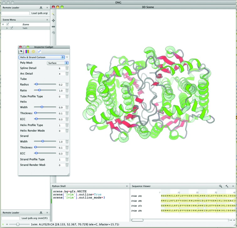
Research projects in structural biology increasingly rely on combinations of heterogeneous sources of information, e.g. evolutionary information from multiple sequence alignments, experimental evidence in the form of density maps and proximity constraints from proteomics experiments. The OpenStructure software framework, which allows the seamless integration of information of different origin, has previously been introduced. The software consists of C++ libraries which are fully accessible from the Python programming language. Additionally, the framework provides a sophisticated graphics module that interactively displays molecular structures and density maps in three dimensions. In this work, the latest developments in the OpenStructure framework are outlined. The extensive capabilities of the framework will be illustrated using short code examples that show how information from molecular-structure coordinates can be combined with sequence data and/or density maps. The framework has been released under the LGPL version 3 license and is available for download from http://www.openstructure.org.

Keywords: OpenStructure; computational structural biology.


Figures

Figure 1
Figure 1
Schematic diagram of the components of entity handles and views. The molecular structure is represented as a tree-like structure rooted at the entity (E). The levels of the tree are formed by chain (C), residue (R) and atom (A). In green, an example entity view containing only a selected subset of elements is shown. The hierarchy of the entity view is separate from the handle; however, at every level the view maps back to its handle, giving access to its properties.
Figure 2
Figure 2
A selection of possible backbone conformations to bridge a fragmented chain. The fragments are coloured by correlation with the density from yellow to green. The tube thickness used to render the backbone fragment is scaled according to the density correlation.
Figure 3
Figure 3
Two distinct visualization styles illustrating the graphical capabilities (see text for a more detailed description). (a) Hemi-light shading with outline mode, (b) simplified enzyme representation by its molecular surface together with an inhibitor.
Figure 4
Figure 4
Visualization of predicted cross-link locations in a homology model of the urease from Y. enterocolitica. The subunits of the urease are coloured blue (α subunits), green (β subunits) and grey (γ subunits).
Figure 5
Figure 5
Screenshot of the graphical user interface DNG. Controls for data display are organized in a main application window. By default, the majority of the main window is taken up by the three-dimensional scene window, which shows a structure rendered in ribbon mode. The user interacts with the scene using the mouse and keyboard shortcuts. On the left side the currently loaded graphical objects are shown in the scene as a tree view that reflects the structure in the scene graph. The render parameters of graphical objects may be changed using the inspector widget displayed on top of the three-dimensional window. In the bottom right corner the sequences of the loaded proteins are shown.

References

    1. Adams, P. D. et al. (2011). Methods, 55, 94–106.
    1. Alber, F., Dokudovskaya, S., Veenhoff, L. M., Zhang, W., Kipper, J., Devos, D., Suprapto, A., Karni-Schmidt, O., Williams, R., Chait, B. T., Sali, A. & Rout, M. P. (2007). Nature (London), 450, 695–701. - PubMed
    1. Altschul, S. F., Gish, W., Miller, W., Myers, E. W. & Lipman, D. J. (1990). J. Mol. Biol. 215, 403–410. - PubMed
    1. Altschul, S. F., Madden, T. L., Schäffer, A. A., Zhang, J., Zhang, Z., Miller, W. & Lipman, D. J. (1997). Nucleic Acids Res. 25, 3389–3402. - PMC - PubMed
    1. Amrein, B., Schmid, M., Collet, G., Cuniasse, P., Gilardoni, F., Seebeck, F. P. & Ward, T. R. (2012). Metallomics, 4, 379–388. - PubMed