

Towards protein-crystal centering using second-harmonic generation (SHG) microscopy
- PMID: 23633594
- PMCID: PMC3640472
- DOI: 10.1107/S0907444913002746
Towards protein-crystal centering using second-harmonic generation (SHG) microscopy
Abstract
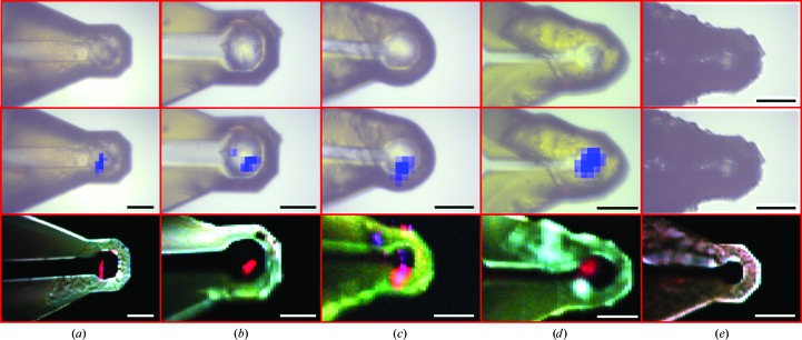
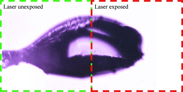
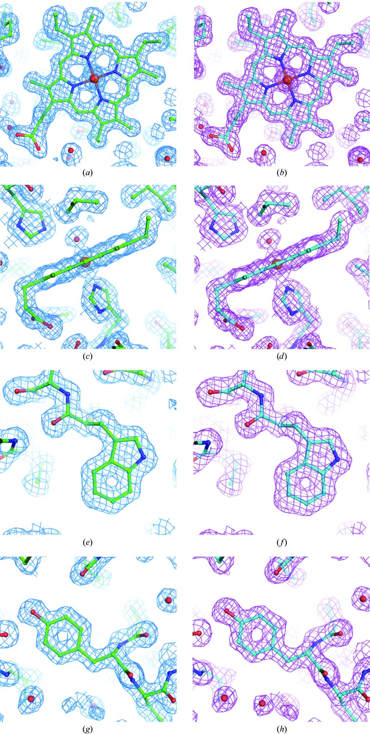
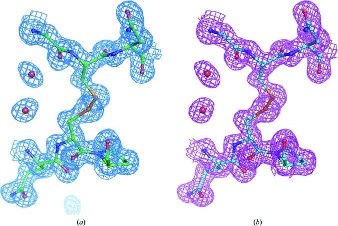
The potential of second-harmonic generation (SHG) microscopy for automated crystal centering to guide synchrotron X-ray diffraction of protein crystals was explored. These studies included (i) comparison of microcrystal positions in cryoloops as determined by SHG imaging and by X-ray diffraction rastering and (ii) X-ray structure determinations of selected proteins to investigate the potential for laser-induced damage from SHG imaging. In studies using β2 adrenergic receptor membrane-protein crystals prepared in lipidic mesophase, the crystal locations identified by SHG images obtained in transmission mode were found to correlate well with the crystal locations identified by raster scanning using an X-ray minibeam. SHG imaging was found to provide about 2 µm spatial resolution and shorter image-acquisition times. The general insensitivity of SHG images to optical scatter enabled the reliable identification of microcrystals within opaque cryocooled lipidic mesophases that were not identified by conventional bright-field imaging. The potential impact of extended exposure of protein crystals to five times a typical imaging dose from an ultrafast laser source was also assessed. Measurements of myoglobin and thaumatin crystals resulted in no statistically significant differences between structures obtained from diffraction data acquired from exposed and unexposed regions of single crystals. Practical constraints for integrating SHG imaging into an active beamline for routine automated crystal centering are discussed.
Keywords: crystal centering; imaging; second-harmonic generation microscopy.
Figures
Similar articles
-
Integrated nonlinear optical imaging microscope for on-axis crystal detection and centering at a synchrotron beamline.J Synchrotron Radiat. 2013 Jul;20(Pt 4):531-40. doi: 10.1107/S0909049513007942. Epub 2013 May 3. J Synchrotron Radiat. 2013. PMID: 23765294 Free PMC article.
-
Dynamic X-ray diffraction sampling for protein crystal positioning.J Synchrotron Radiat. 2017 Jan 1;24(Pt 1):188-195. doi: 10.1107/S160057751601612X. Epub 2017 Jan 1. J Synchrotron Radiat. 2017. PMID: 28009558 Free PMC article.
-
Goniometer-based femtosecond crystallography with X-ray free electron lasers.Proc Natl Acad Sci U S A. 2014 Dec 2;111(48):17122-7. doi: 10.1073/pnas.1418733111. Epub 2014 Oct 31. Proc Natl Acad Sci U S A. 2014. PMID: 25362050 Free PMC article.
-
Rastering strategy for screening and centring of microcrystal samples of human membrane proteins with a sub-10 microm size X-ray synchrotron beam.J R Soc Interface. 2009 Oct 6;6 Suppl 5(Suppl 5):S587-97. doi: 10.1098/rsif.2009.0142.focus. Epub 2009 Jun 17. J R Soc Interface. 2009. PMID: 19535414 Free PMC article. Review.
-
Collection of X-Ray Diffraction Data from Macromolecular Crystals.Methods Mol Biol. 2017;1607:165-184. doi: 10.1007/978-1-4939-7000-1_7. Methods Mol Biol. 2017. PMID: 28573573 Free PMC article. Review.
Cited by
-
Integrated nonlinear optical imaging microscope for on-axis crystal detection and centering at a synchrotron beamline.J Synchrotron Radiat. 2013 Jul;20(Pt 4):531-40. doi: 10.1107/S0909049513007942. Epub 2013 May 3. J Synchrotron Radiat. 2013. PMID: 23765294 Free PMC article.
-
Approaches to automated protein crystal harvesting.Acta Crystallogr F Struct Biol Commun. 2014 Feb;70(Pt 2):133-55. doi: 10.1107/S2053230X14000387. Epub 2014 Jan 28. Acta Crystallogr F Struct Biol Commun. 2014. PMID: 24637746 Free PMC article.
-
The complex analysis of X-ray mesh scans for macromolecular crystallography.Acta Crystallogr D Struct Biol. 2018 Apr 1;74(Pt 4):355-365. doi: 10.1107/S2059798318002735. Epub 2018 Apr 6. Acta Crystallogr D Struct Biol. 2018. PMID: 29652262 Free PMC article.
-
Kinetic trapping of metastable amino acid polymorphs.J Am Chem Soc. 2014 Feb 12;136(6):2404-12. doi: 10.1021/ja410293p. Epub 2014 Feb 4. J Am Chem Soc. 2014. PMID: 24451055 Free PMC article.
-
Strategies for sample delivery for femtosecond crystallography.Acta Crystallogr D Struct Biol. 2019 Feb 1;75(Pt 2):160-177. doi: 10.1107/S2059798318017953. Epub 2019 Feb 19. Acta Crystallogr D Struct Biol. 2019. PMID: 30821705 Free PMC article. Review.
References
-
- Asanov, A. N., McDonald, H. M., Oldham, P. B., Jedrzejas, M. J. & Wilson, W. (2001). J. Cryst. Growth, 232, 603–609.
-
- Asherie, N., Jakoncic, J., Ginsberg, C., Greenbaum, A., Stojanoff, V., Hrnjez, B. J., Blass, S. & Berger, J. (2009). Cryst. Growth Des. 9, 4189–4198.
-
- Beitlich, T., Kühnel, K., Schulze-Briese, C., Shoeman, R. L. & Schlichting, I. (2007). J. Synchrotron Rad. 14, 11–23. - PubMed
-
- Bourgeois, D., Vernede, X., Adam, V., Fioravanti, E. & Ursby, T. (2002). J. Appl. Cryst. 35, 319–326.
Publication types
MeSH terms
Substances
Associated data
- Actions
- Actions
- Actions
- Actions
Grants and funding
LinkOut - more resources
Full Text Sources
Other Literature Sources
