

Ultrasound-guided percutaneous antegrade pyelography with computed tomography for the diagnosis of spontaneous partial ureteral rupture in a dog
- PMID: 23633712
- PMCID: PMC3474574
Ultrasound-guided percutaneous antegrade pyelography with computed tomography for the diagnosis of spontaneous partial ureteral rupture in a dog
Abstract
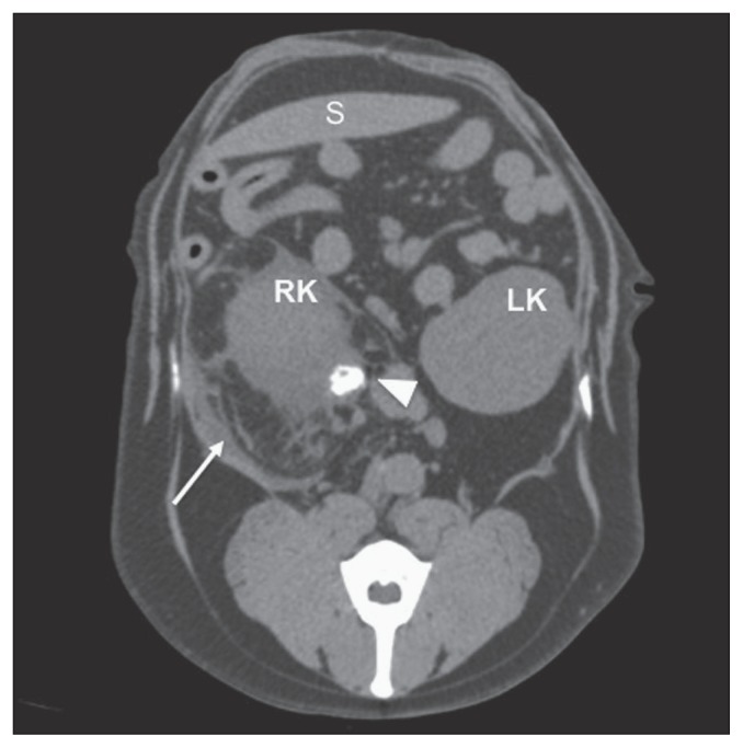
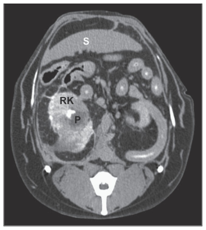
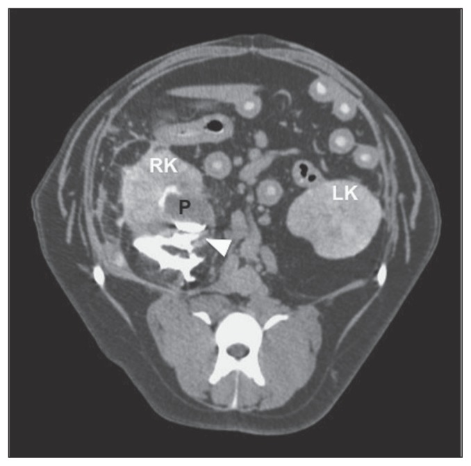
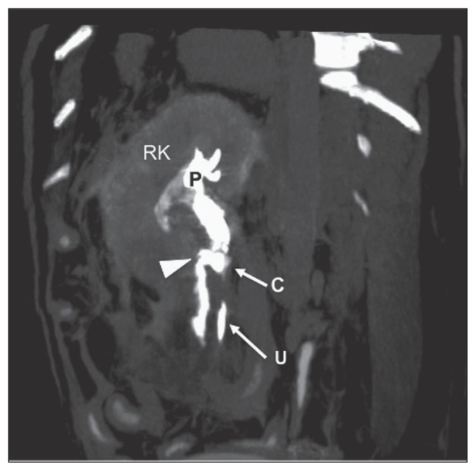
A 10-year-old spayed female dalmatian dog developed acute vomiting and abdominal pain. Ultrasound examination of the abdomen showed right hydronephrosis and proximal ureter dilation with mild retroperitoneal free fluid. Computed tomography (CT) of the abdomen confirmed the ultrasonographic findings and revealed, additionally, a right ureteral stone. Spontaneus rupture of the right ureter was confirmed with CT post ultrasound-guided percutaneous antegrade pyelography. Pyeloureteral rupture and the presence of a ureteral stone were confirmed at surgery.
Pyélographie antégrade percutanée guidée par échographie avec tomodensitométrie pour le diagnostic d’une rupture urétrale partielle spontanée chez un chien. Une chienne Dalmatien stérilisée âgée de 10 ans a manifesté des vomissements et de la douleur abdominale aigus. Une échographie de l’abdomen a montré de l’hydronéphrose à droite et une dilatation proximale de l’urètre avec un peu de liquide rétropéritonéal libre. Une tomodensitométrie de l’abdomen a confirmé les résultats de l’échographie et a révélé, en plus, un calcul urétéral droit. Une rupture spontanée de l’urètre droit a été confirmée par tomodensitométrie après une pyélographie antégrade percutanée guidée par échographie. La rupture pyélo-urétérale et la présence de calcul urétéral ont été confirmées à la chirurgie.(Traduit par Isabelle Vallières).
Figures
References
-
- Gayer G, Zissin R, Apter S, et al. Urinomas caused by ureteral injuries: CT appearance. Abdom Imaging. 2002;27:88–92. - PubMed
-
- Hanika C, Rebar AH. Ureteral transitional cell carcinoma in the dog. Vet Pathol. 1980;17:643–646. - PubMed
-
- Kyles AE, Douglas JP, Rottman JB. Pyelonephritis following inadvertent excision of the ureter during ovariohysterectomy in a bitch. Vet Rec. 1996;139:471. - PubMed
-
- Weisse C, Aronson LR, Drobatz KJ. Traumatic rupture of the ureter: 10 cases. J Am Anim Hosp Assoc. 2002;38:188–194. - PubMed
-
- Jackman SV, Potter SR, Regan F, Jarrett TW. Plain abdominal x-ray versus computerized tomography screening: Sensitivity for stone localization after nonenhanced spiral computerized tomography. J Urol. 2000;164:308–310. - PubMed
Publication types
MeSH terms
LinkOut - more resources
Full Text Sources
Medical