

Oral myiasis in an adult associated with filariasis and Hansen's disease
- PMID: 23633879
- PMCID: PMC3633295
- DOI: 10.4103/0976-9668.107322
Oral myiasis in an adult associated with filariasis and Hansen's disease
Abstract
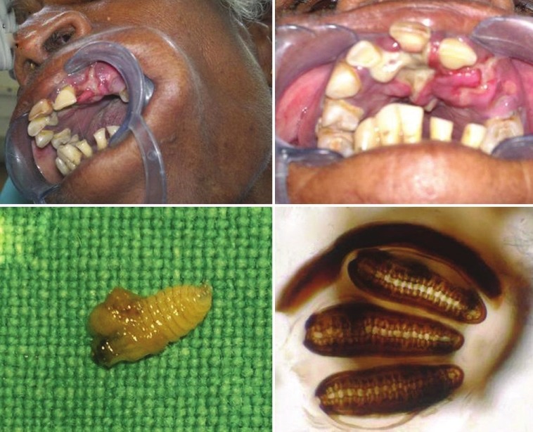
Oral myiasis is a common parasitic infestation of live human and animals caused by species of dipteran fly larvae known as maggots which may be secondary to medical disease. This case involves a 51-year-old female, poorly debilitated with advanced periodontal disease infected by the dipteral larvae in the anterior maxillary region which belonged to the family Calliphoridae and Chrysomya bezziana species. This lady was neglected from her family and presented oral myiasis with the previous history of filariasis and Hansen's disease. Secondary infestations may occur in cancrum oris, oral extraction wounds, jaw bone wounds, oral leprosy lesion, filariasis, and carcinoma. Hansen's disease (leprosy) is bacterial in origin whereas filariasis (elephantiasis) is parasitic in origin like-myiasis. The treatment consisted of manual removal of the larvae by topical application of turpentine oil, oral therapy, and surgical debridement of the oral wound.
Keywords: Filariasis; Hansen's disease; flies larvae; maggots; oral myiasis.
Conflict of interest statement
Figures


Similar articles
-
Management of Oral Myiasis Caused by Chrysomya bezziana - A Case Series.Ann Maxillofac Surg. 2020 Jul-Dec;10(2):521-524. doi: 10.4103/ams.ams_177_20. Epub 2020 Dec 23. Ann Maxillofac Surg. 2020. PMID: 33708609 Free PMC article.
-
Primary oral myiasis: a case report.Med Oral Patol Oral Cir Bucal. 2008 Nov 1;13(11):E714-6. Med Oral Patol Oral Cir Bucal. 2008. PMID: 18978712
-
Oral myiasis: case report.Indian J Dent Res. 2013 Nov-Dec;24(6):750-2. doi: 10.4103/0970-9290.127626. Indian J Dent Res. 2013. PMID: 24552939
-
Cryptic Myiasis by Chrysomya bezziana: A Case Report and Literature Review.Turk J Ophthalmol. 2020 Dec 29;50(6):381-386. doi: 10.4274/tjo.galenos.2020.69360. Turk J Ophthalmol. 2020. PMID: 33389940 Free PMC article. Review.
-
Cutaneous myiasis: a review of the common types of myiasis.Int J Dermatol. 2010 Oct;49(10):1092-8. doi: 10.1111/j.1365-4632.2010.04577.x. Int J Dermatol. 2010. PMID: 20883399 Review.
Cited by
-
Oral Myiasis Caused by Chrysomya bezziana in Anterior Maxilla.Case Rep Dent. 2014;2014:518427. doi: 10.1155/2014/518427. Epub 2014 Apr 29. Case Rep Dent. 2014. PMID: 24872898 Free PMC article.
-
Management of Oral Myiasis Caused by Chrysomya bezziana - A Case Series.Ann Maxillofac Surg. 2020 Jul-Dec;10(2):521-524. doi: 10.4103/ams.ams_177_20. Epub 2020 Dec 23. Ann Maxillofac Surg. 2020. PMID: 33708609 Free PMC article.
-
Oral Myiasis: A Rare Case Report and Literature Review.J Dent (Tehran). 2015 Jun;12(6):456-9. J Dent (Tehran). 2015. PMID: 26884780 Free PMC article.
References
-
- Laurence SM. Dipterous larvae infection. Br Med J. 1909;9:88.
-
- Sheikh S, Pallagatti S, Singla I, Kalucha A, Aggarwal A, Kaur H. Oral Myiasis: A review. J Clin Exp Dent. 2011;3:e465–8.
-
- Thami GP, Baurah MC, Sharmce SC, Behera NK. Nasal myiasis in leprosy leading to unusual tissue destruction. J Dermatol. 1995;22:348–50. - PubMed
Publication types
LinkOut - more resources
Full Text Sources
Other Literature Sources