. 2013;9(4):e1003325.
doi: 10.1371/journal.ppat.1003325. Epub 2013 Apr 25.

The Chlamydia pneumoniae invasin protein Pmp21 recruits the EGF receptor for host cell entry

Affiliations

The Chlamydia pneumoniae invasin protein Pmp21 recruits the EGF receptor for host cell entry

Katja Mölleken et al. PLoS Pathog. 2013.

Abstract

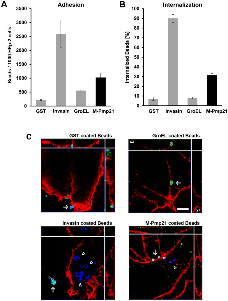
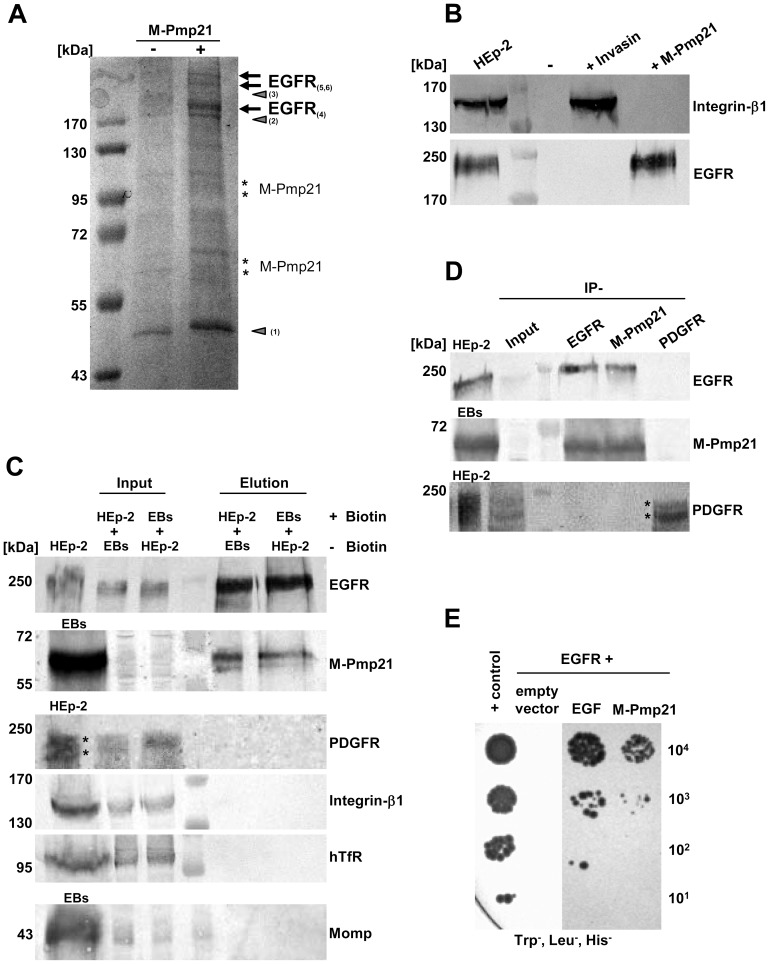
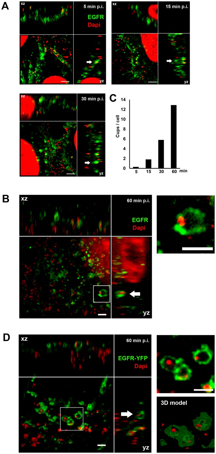
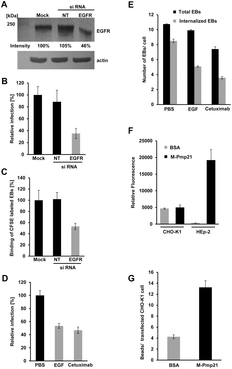
Infection of mammalian cells by the strictly intracellular pathogens Chlamydiae requires adhesion and internalization of the infectious Elementary Bodies (EBs). The components of the latter step were unknown. Here, we identify Chlamydia pneumoniae Pmp21 as an invasin and EGFR as its receptor. Modulation of EGFR surface expression evokes correlated changes in EB adhesion, internalization and infectivity. Ectopic expression of EGFR in EGFR-negative hamster cells leads to binding of Pmp21 beads and EBs, thus boosting the infection. EB/Pmp21 binding and invasion of epithelial cells results in activation of EGFR, recruitment of adaptors Grb2 and c-Cbl and activation of ERK1/2, while inhibition of EGFR or MEK kinase activity abrogates EB entry, but not attachment. Binding of Grb2 and c-Cbl by EGFR is essential for infection. This is the first report of an invasin-receptor interaction involved in host-cell invasion by any chlamydial species.


Conflict of interest statement

The authors have declared that no competing interests exist.

Figures

Figure 1
Figure 1. Pmp21-coated beads are taken up by mammalian cells.
(A) Green fluorescent beads coated with recombinant GST, invasin, GroEL1 or M-Pmp21 were incubated in 5-fold excess with HEp-2 cells for 1 h at 4°C, and the numbers of beads found on 1000 HEp-2 cells were counted (n = 3). (B) Internalization of M-Pmp21-coated beads. HEp-2 cells were incubated with beads as above at 37°C for 4 h, and washed with PBS to remove unattached beads. Attached beads were stained with specific antibodies without cell permeabilization (see Figure S1), and the numbers of attached (red) and internalized (green) beads on/in samples of 1000 cells were counted (n = 3). (C) Confocal spinning-disk images of internalized beads (for the GroEL image the xz and yz plain projections of the whole field are marked). HEp-2 cells were incubated for 4 h at 37°C with blue fluorescent beads coated with GST, invasin, GroEL1 or M-Pmp21. External beads were stained with protein specific antibodies (green). Internalized beads are not accessible to the antibody and appear blue. Cell boundaries were stained with Wheat Germ Agglutinin-Alexa594 (red). External beads are marked by white arrows, internalized beads by white triangles. Bar 5 µm.
Figure 2
Figure 2. Identification of the EGF receptor as an interaction partner for the C. pneumoniae adhesin Pmp21.
(A) Electrophoretic analysis of fractions eluted from a NeutrAvidin column. HEp-2 cells were incubated with (+) or without (−) biotinylated M-Pmp21 or invasin (data for invasin not shown), and processed as described in Experimental Procedures. Arrows mark bands in which EGFR was identified by MALDI-MS (see Fig. S2B; bands 4–6). Other major bands (triangles) were identified as actin (1) and pleckstrin (2); no protein was detected in band 3. Bands marked with asterisks represent the recombinant M-Pmp21 as shown by immunoblotting (data not shown). (B) Fractions from (A) were probed with anti-integrin-β1 and anti-EGFR antibodies. (C) Affinity purification of the EGFR-Pmp21 complex from surface-biotinylated HEp-2 cells incubated with non-biotinylated C. pneumoniae EBs, or vice versa, for 60 min at 37°C. After crosslinking the biotinylated surface protein complexes were bound to a NeutrAvidin column. After crosslink removal the eluted proteins were identified by immunoblot analysis with specific antibodies against EGFR, M-Pmp21, PDGFR, integrin-β1, hTfR and Momp. Equal amounts of input and elution samples were loaded onto the SDS-PAGE. (D) Coimmunoprecipitation of the EGFR-Pmp21 complex from HEp-2 monolayers incubated with purified C. pneumoniae EBs (MOI 5) for 60 min. The Pmp21-EGFR complex was precipitated using EGFR-, M-Pmp21- and PDGFR-specific antibodies as indicated, and probed after SDS-PAGE and immunoblotting. Stars mark the 2 main PDGFR protein species recognized. (E) Interaction of EGFR and M-Pmp21 shown by Y2H analysis. Serial dilutions (101–104) of yeast cells expressing EGFR/EGF or EGFR/M-Pmp21 were patched on low-stringency selection medium (Leu, Trp, His). + control: SV40 LTA/p53.
Figure 3
Figure 3. EGFR colocalizes with EBs at bacterial entry sites.
(A–B) Confocal spinning-disk immunofluorescence microscopy of HEp-2 cells exposed to C. pneumoniae EBs for 5 min, 15 min, 30 min (A) or 60 min (B). Fixed cells were stained with anti-EGFR (green) and EBs with DAPI (red). Arrows indicate colocalization of the two signals. (B) C. pneumoniae EBs surrounded by ring-like EGFR structures 60 min pi. Arrows indicate colocalization of EGFR with DAPI-stained EBs at bacterial entry sites in xz and yz planes. An internalized EB surrounded by EGFR (white box) is shown at higher magnification on the right. (C) Quantification of colocalization events (cups) of EBs and EGFR signals at the indicated time points. The data are from a typical experiment. (D) Transfected CHO-K1 cells expressing human EGFR tagged with YFP (green) reveal colocalization of EGFR with DAPI-stained EBs (red) at bacterial entry sites 60 min pi. An internalized EB surrounded by EGFR-YFP (white box) is shown at a higher magnification on the upper right, and a 3D model of the structure (based on a Maximal Intensity Projection) is shown below it (see movie S1). Bar 1 µm.
Figure 4
Figure 4. EGFR is required for internalization of C. pneumoniae.
(A–C) HeLa229 cells were mock-transfected (Mock), or transfected with a non-targeting siRNA (NT) or an EGFR-targeting siRNA for 24 h. (A) Relative EGFR levels were determined using Scion Image software after immunoblot analysis with anti-EGFR. Actin served as loading control. (B) HeLa229 cells transfected for 24 h were infected with C. pneumoniae GiD (MOI 1) for 48 h. Inclusion formation was evaluated by indirect immunofluorescence with an FITC-conjugated antibody against chlamydial LPS. The number of inclusions in mock-transfected cells was set to 100% (n = 4). (C) C. pneumoniae EBs labeled with CFSE were added (MOI 10) to transfected HeLa229 cells for 1 h at 37°C. Cells were then detached from the substrate and fixed with formaldehyde, and adhesion was measured by flow cytometry. The mean fluorescence of EBs bound to mock-transfected cells was set to 100% (n = 3). (D) Pretreatment of confluent HEp-2 monolayers with recombinant EGF or Cetuximab for 2 h inhibits subsequent infection (n = 4) (MOI 1). (E) Pretreatment of HEp-2 cells with rEGF or Cetuximab for 2 h inhibits subsequent internalization of C. pneumoniae EBs (MOI 1). Cells were exposed to EBs for 2 h, then fixed and stained with anti-Pmp21 and DAPI without permeabilization. Numbers of internalized EBs (inaccessible to anti-Pmp21 antibody) were determined by subtracting the number of external EBs (visualized with anti-Pmp21) from the total number of EBs (DAPI stain) in each cell (n = 5). (F) rM-Pmp21-coated beads adhere to HEp-2 but not CHO-K1 cells. A five-fold excess of green fluorescent beads coupled to BSA or rM-Pmp21 were incubated with CHO-K1 and HEp-2 cells for 1 h at 37°C. Unbound beads were removed by washing with PBS, and cells bearing attached beads were analyzed by flow cytometry. The mean fluorescence values for the samples analyzed (n = 4) are shown. (G) rM-Pmp21-coated beads attach to EGFR-expressing CHO-K1 cells. CHO-K1 cells were transfected with EGFR-mCherry for 24 h, incubated with a five-fold excess of green fluorescent beads coated with BSA or rM-Pmp21, and the numbers of beads attached to transfected cells were determined (n = 3).
Figure 5
Figure 5. EGFR signaling is activated by C. pneumoniae EBs and recombinant Pmp21.
(A) Kinetics of C. pneumoniae EB-induced phosphorylation of EGFR. HEp-2 cells were left uninfected (0) or infected with increasing numbers of C. pneumoniae EBs (MOI) for 60 min. The immunoblots show total levels of EGFR (EGFR) and levels of activated receptor (pEGFR, phosphorylated at Tyr1068) detected as described in Experimental Procedures. (B) Time courses of EGFR activation by rEGF (100 ng/ml), purified (viable and non-viable) C. pn. EBs (MOI 5), rM-Pmp21 (100 µg/ml) in the presence or absence of Cetuximab (5 µg/ml), and rOmcB (100 µg/ml). HEp-2 cells were serum-starved for 12 h at 37°C, shifted to 4°C for 10 min before the addition of bacteria or recombinant proteins, then incubated further at 37°C. The immunoblots show overall levels of EGFR (EGFR), activated EGFR (pEGFR) and activated ERK (pERK), the downstream MAP kinase. Actin served as loading control. (C–D) Confocal spinning-disk microscopy of C. pneumoniae EBs (C) or rM-Pmp21-coated latex beads (D) surrounded by ring-like structures of activated EGFR (pEGFRY1068). Scale bars 1 µm. (C) HEp-2 cells were infected with C. pneumoniae for 60 min. Two examples of internalized EBs surrounded by EGFR (white boxes) are shown at a higher magnification on the right. (D) Beads coated with rM-Pmp21 were added to EGFR-YFP-expressing CHO-K1 cells and incubated for 4 h at 37°C. The arrow marks a bead in the focal plane harboring EGFR and pEGFR rings. (E) EGFR inhibitors reduce infection by C. pneumoniae EBs. Confluent HEp-2 cells were pretreated for 2 h with PBS, AG1478 or UO126, and infected with C. pneumoniae EBs (MOI 1) for 48 h. The data represent the means of four independent experiments. (F) Internalization of C. pneumoniae EBs (MOI 1) by HEp-2 cells treated for 2 h with PBS, AG1478 or UO126. Internalized EBs were quantified as described in Experimental Procedures. The data represent the means of five independent experiments.
Figure 6
Figure 6. A functional EGF-binding domain in EGFR is essential for infection by C. pneumoniae.
(A–D) EGFR-deficient CHO-K1 cells were transfected with EGFR-YFP, EGFRΔBD2-YFP or YFP alone for 24 h. (A) EGFR expression was quantified by immunoblot analysis of lysates of transfected cells using an anti-EGFR antibody. (B) Subcellular localization of YFP and the two EGFR-YFP constructs by direct immunofluorescence of transfected CHO-K1 cells. Bar 5 µm. (C) Susceptibility of transfected CHO-K1 cells to infection with C. pneumoniae GiD. Cells were incubated with EBs (MOI 1) for 48 h. Inclusions were quantified using an antibody directed against the inclusion membrane protein Cpn0147. The data represent the means of four independent experiments. (D) Internalization of C. pneumoniae EBs (MOI 1) by CHO-K1 cells transfected with YFP, EGFR-YFP or EGFRΔBD2-YFP. Numbers of internalized EBs were determined in positively transfected cells only. The data represent the means of five independent experiments. (E) Y2H analysis of EGFRΔBD2/EGF and EGFRΔBD2/M-Pmp21 interactions. Serial dilution patch test of 101–104 cells on selective medium.
Figure 7
Figure 7. The EGFR adaptor proteins c-Cbl and Grb2 colocalize with invading C. pneumoniae EBs.
(A) c-Cbl and Grb2 specifically interact with invading C. pneumoniae EBs. Adaptor proteins c-Cbl and Grb2 were affinity-enriched from surface-biotinylated HEp-2 cells incubated with non-biotinylated C. pneumoniae EBs, or vice versa, for 60 min at 37°C. The eluted proteins were subjected to immunoblot analysis with specific antibodies against c-Cbl, Grb2 or PDGFRβ. Equal amounts of input and elution samples were loaded onto the SDS/PAGE. (B–C) c-Cbl and Grb2 colocalize with EGFR and invading EBs. (B) Confocal spinning-disk microscopy of C. pneumoniae EBs infecting HEp-2 cells for 60 min at 37°C. The arrow marks a single DAPI-stained EB (blue) colocalizing with antibody-stained endogenous EGFR (green) and endogenous c-Cbl (red) or endogenous Grb2 (red). Bar 1 µm. Below each picture an intensity scan along the indicated white line is shown (blue – DAPI signal; green – EGFR signal; red – c-Cbl or Grb2 signal). Quantification of colocalization of EBs stained by DAPI and c-Cbl or Grb2 signals is shown below. The data represent the means of five independent experiments. (C) Colocalization of transfected EGFR-mCherry with either c-Cbl-YFP or Grb2-YFP and invading C. pneumoniae EBs in CHO-K1 cells. The arrow marks a single EB surrounded by an EGFR-mCherry signal and either c-Cbl-YFP or Grb2-YFP signals. Bar 1 µm. (D) Functional interaction between EGFR and c-Cbl and Grb2 is essential for infection by C. pneumoniae. CHO-K1 cells were transfected with wt EGFR or the mutant EGFRY1045F (docking site for c-Cbl) or EGFRY1068/1086F (docking site for Grb2) and 24 h later infected with C. pneumoniae for 48 h. Inclusions were quantified using an antibody against Cpn0147 (n = 4). (E) Internalization of C. pneumoniae EBs (MOI 1) by CHO-K1 cells transfected with EGFR-YFP or EGFRY1045F or EGFRY1068/1086F. Numbers of internalized EBs were determined in positively transfected cells only. The data represent the means of five independent experiments.

References

    1. Campbell LA, Kuo CC (2004) Chlamydia pneumoniae–an infectious risk factor for atherosclerosis? Nat Rev Microbiol 2: 23–32. - PubMed
    1. Boleti H, Benmerah A, Ojcius DM, Cerf-Bensussan N, Dautry-Varsat A (1999) Chlamydia infection of epithelial cells expressing dynamin and Eps15 mutants: clathrin-independent entry into cells and dynamin-dependent productive growth. J Cell Sci 112: 1487–1496. - PubMed
    1. Hybiske K, Stephens RS (2007) Mechanisms of Chlamydia trachomatis Entry into Nonphagocytic Cells. Infect Immun 75: 3925–3934. - PMC - PubMed
    1. Dautry-Varsat A, Subtil A, Hackstadt T (2005) Recent insights into the mechanisms of Chlamydia entry. Cell Microbiol 7: 1714–1722. - PubMed
    1. Carabeo RA, Grieshaber SS, Fischer E, Hackstadt T (2002) Chlamydia trachomatis induces remodeling of the actin cytoskeleton during attachment and entry into HeLa cells. Infect Immun 70: 3793–3803. - PMC - PubMed

Publication types

MeSH terms

Substances