. 2013 Apr 25;8(4):e61925.
doi: 10.1371/journal.pone.0061925. Print 2013.

Inhibition of nuclear factor-kappa B activation decreases survival of Mycobacterium tuberculosis in human macrophages

Affiliations

Inhibition of nuclear factor-kappa B activation decreases survival of Mycobacterium tuberculosis in human macrophages

Xiyuan Bai et al. PLoS One. .

Abstract

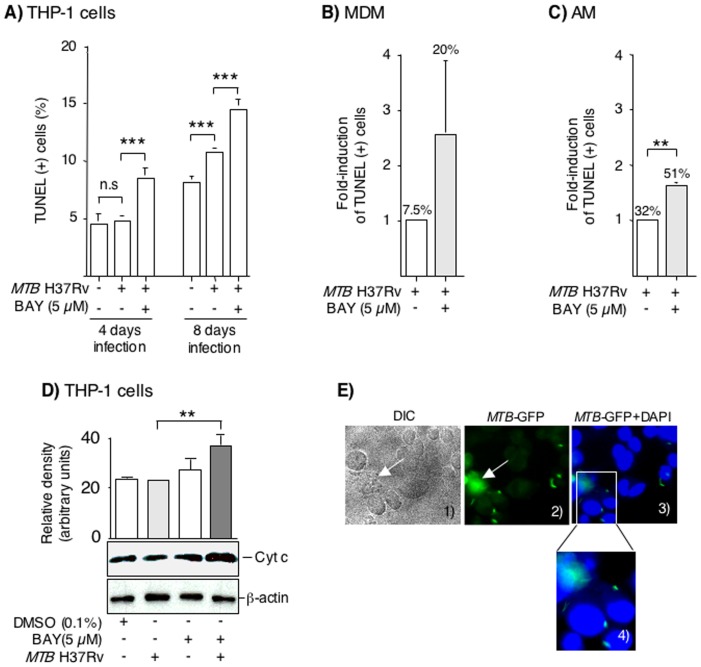
Nuclear factor-kappa B (NFκB) is a ubiquitous transcription factor that mediates pro-inflammatory responses required for host control of many microbial pathogens; on the other hand, NFκB has been implicated in the pathogenesis of other inflammatory and infectious diseases. Mice with genetic disruption of the p50 subunit of NFκB are more likely to succumb to Mycobacterium tuberculosis (MTB). However, the role of NFκB in host defense in humans is not fully understood. We sought to examine the role of NFκB activation in the immune response of human macrophages to MTB. Targeted pharmacologic inhibition of NFκB activation using BAY 11-7082 (BAY, an inhibitor of IκBα kinase) or an adenovirus construct with a dominant-negative IκBα significantly decreased the number of viable intracellular mycobacteria recovered from THP-1 macrophages four and eight days after infection. The results with BAY were confirmed in primary human monocyte-derived macrophages and alveolar macrophages. NFκB inhibition was associated with increased macrophage apoptosis and autophagy, which are well-established killing mechanisms of intracellular MTB. Inhibition of the executioner protease caspase-3 or of the autophagic pathway significantly abrogated the effects of BAY. We conclude that NFκB inhibition decreases viability of intracellular MTB in human macrophages via induction of apoptosis and autophagy.


Conflict of interest statement

Competing Interests: The Authors have declared that no competing interests exist.

Figures

Figure 1
Figure 1. MTB H37Rv induces NFκB binding to its cis-regulatory element.
(A) THP-1 cells were pre-treated with 0.1% (v/v) DMSO or 5 µM BAY for 1 hr, followed by infection with MTB H37Rv at the indicated times. An EMSA was performed with an oligonucleotide that corresponds to the consensus binding sequence for NFκB. Data shown are representative of three independent experiments. (B) Primary human MDM or (C) AM were pre-incubated with 0.1% DMSO or 5 µM BAY for 1 hr, then infected with MTB for 3 hrs, and followed by an EMSA to assay for NFκB binding. Data shown are representative of two independent experiments for MDM and AM. NS = non-specific band. SS = supershift band.
Figure 2
Figure 2. Inhibition of NFκB activation by BAY 11-7082 reduces the viability of intracellular MTB in human macrophages.
(A) THP-1 cells, (B) MDM, or (C) AM were pretreated with 0.1% (v/v) DMSO (open squares) or 5 µM BAY (closed circles) for 1 hr, followed by infection with MTB H37Rv. One hr, 4 days, or 8 days after infection, the cells were lysed and cultured for MTB. (D) Differentiated THP-1 cells were pretreated with 0.1% (v/v) DMSO (open squares) or 5 µM BAY (closed circles) for one 1 hr, followed by infection with MTB-H37Rv-GFP. One hr, 4 days, or 8 days after infection, fluorescent intensity was measured by Cytofluor II microplate fluorometer. Data shown as mean ± SEM. n = 4 for THP-1 cells in (A) and n = 2 for THP-1 cells in (D), n = 7 volunteers for MDM, n = 9 volunteers for AM. *p<0.05, **p<0.01, ***p<0.001.
Figure 3
Figure 3. Inhibition of NFκB activation using a dominant-negative to IκBα.
(A) THP-1 cells were transduced with or without AdV-GFP or AdV-S32/36A-IκBα at a MOI of 30∶1 for 5 hrs and then infected with MTB H37Rv. After 4 days of infection, the cells were lysed and cell-associated MTB quantified. (B) Wildtype THP-1 cells with or without pre-treatment with 5 µM BAY were infected with MTB H37Rv. Other THP-1 cells were transduced with AdV-GFP or AdV-S32/36A-IκBα at a MOI of 30∶1 for 5 hrs and then infected with MTB H37Rv. After 24 hrs of infection, the supernatants were measured for IL-8 by ELISA. Data are means ± SEM of two independent experiments performed in duplicates. *p<0.05, ** p<0.01.
Figure 4
Figure 4. Inhibition of NFκB activation increases apoptosis of infected macrophages.
(A) THP-1 cells were infected with MTB H37Rv for 4 and 8 days with or without BAY, and apoptosis measured by TUNEL. Data for THP-1 cells are the mean ± SEM of four independent experiments. (B) Primary human MDM and (C) AM were infected with MTB with or without BAY, cultured for 4 days, and apoptosis measured by TUNEL. The percentage (%) numbers above the bars indicate the % cells with positive TUNEL stain. Data for MDM and AM are the mean ± SEM of three independent experiments. n.s. = not significant, **p<0.01, ***p<0.001. (D) THP-1 cells were infected with MTB, 5 µM BAY 11-7082, or both. After 48 hrs, nuclear-free whole cell lysates isolated, and western blot performed for cytochrome c. The membranes were also immunoblotted for β-actin. The bar graph above the immunoblot represent the mean relative density measurements for cytochrome c bands normalized for the densities of the corresponding β-actin band. The data shown are representative of two independent experiments. **p<0.01. (E) THP-1 cells were infected with MTB H37Rv-GFP for 1 hr, stained with DAPI, and viewed under both differential interference contrast (DIC) and fluorescent imaging under 630× magnification (panels 1–3). An area of panel 3 was magnified further on the computer screen (panel 4). Data shown are representative of two independent experiments.
Figure 5
Figure 5. Inhibition of caspase-3 activation abrogates the effects of BAY.
(A) THP-1 cells were cultured with 5 µM BAY, MTB, or MTB+BAY with or without 10 µM of the caspase-3 inhibitor z-DEVD-fmk for 48 hrs. After the indicated time, the cells were lysed and activated caspase-3 quantified by ELISA. Data shown are the mean ± SEM of two independent experiments performed in duplicates. *p<0.05, **p<0.01. (B) THP-1 cells were infected with MTB H37Rv alone (open diamonds), MTB+BAY (closed squares), or MTB+BAY+z-DEVD-fmk (semi-closed triangles). One hr, 2 days, and 4 days after infection, THP-1 cells were lysed and cultured for MTB. (C) The same treatment conditions as in (A) were repeated with THP-1 cells for 2 days followed by measurement of apoptosis using TUNEL staining. Data shown are the mean ± SEM of two independent experiments. *p<0.05, **p<0.01 and ***p<0.001.
Figure 6
Figure 6. Inhibition of NFκB activation induces autophagy in THP-1 cells.
(A) Control THP-1 cells (0.1% DMSO) and THP-1 cells subjected to serum starvation, 5 µM BAY, MTB infection, or both MTB+BAY for 24 hrs, followed by immunoblotting of nuclear-free whole cell lysates for LC3 and β-actin. A representative immunoblot of three independent experiments is shown. (B) Human THP-1 cells were transduced with lentivirus-GFP-LC3 and differentiated into macrophages, followed by infection with MTB for 24 hrs in the absence or presence of 5 µM BAY. The cells were fixed and stained with DAPI to visualize the nuclei (blue) and the number of GFP-positive punctae were quantified. Upper panel, representative immunofluorescence images of three independent experiments; lower panel, average number of GFP-LC3 punctae per cell. The data shown represent the mean ± SEM of duplicate wells/condition from three independent experiments. (C) MTB-infected THP-1 cells treated with BAY were incubated with or without 3-MA, an inhibitor of the early phase of the autophagic pathway. After 48 hrs, the cells were lysed and nuclear-free whole cell lysates (20 µg per lane) were separated by SDS-PAGE and immunoblotted for LC3-I, LC3-II and β-actin. The bar graph represents the relative densities of the LC3-II bands normalized for their corresponding β-actin bands for two independent experiments. (D) THP-1 cells were infected with MTB H37Rv alone, MTB+5 µM BAY, or MTB+BAY+6 mM 3-MA for 4 days and cell-associated MTB was quantified. Data shown are mean ± SEM from two independent experiments performed in duplicates. *p<0.05, **p<0.01, ***p<0.001.
Figure 7
Figure 7. TNFα and IFNγ levels in MDM and AM infected with MTB H37Rv with or without NFκB inhibition.
Primary human (A) MDM or (B) AM were infected with MTB H37Rv and after 1 hr, 24 hrs, 4 days, or 8 days of infection, supernatants were assayed for TNFα by ELISA and IFNγ by electrochemiluminescence. Data shown are estimated means with standard error bars from linear mixed model fits, based on seven independent experiments (MDM) or nine independent experiments (AM). *p<0.05 and **p<0.01 for cytokine expression in macrophages infected with MTB alone (closed circles) vs. MTB+BAY (closed squares). Control = (open circles) and BAY = (open squares).
Figure 8
Figure 8. Diagram of the mechanisms by which NFκB activation promotes the intracellular survival of MTB.
Based on our experimental findings, NFκB activation enhanced the intracellular survival of MTB through inhibition of apoptosis and autophagy in infected macrophages. Since NFκB can also induce the production of its inhibiting molecule IκBα (blue line) and NFκB inhibition of autophagy could potentially prevent degradation of IKK (red line), the ultimate effect of NFκB on survival of intracellular MTB in macrophages is likely a complex process. IKK = IκBα kinase.

Similar articles

Cited by

  • Autophagy in Mycobacterium tuberculosis and HIV infections.
    Espert L, Beaumelle B, Vergne I. Espert L, et al. Front Cell Infect Microbiol. 2015 Jun 2;5:49. doi: 10.3389/fcimb.2015.00049. eCollection 2015. Front Cell Infect Microbiol. 2015. PMID: 26082897 Free PMC article. Review.
  • Antibiotic adjuvants: synergistic tool to combat multi-drug resistant pathogens.
    Kumar V, Yasmeen N, Pandey A, Ahmad Chaudhary A, Alawam AS, Ahmad Rudayni H, Islam A, Lakhawat SS, Sharma PK, Shahid M. Kumar V, et al. Front Cell Infect Microbiol. 2023 Dec 20;13:1293633. doi: 10.3389/fcimb.2023.1293633. eCollection 2023. Front Cell Infect Microbiol. 2023. PMID: 38179424 Free PMC article. Review.
  • BCG vaccination reduces the mortality of Mycobacterium tuberculosis-infected type 2 diabetes mellitus mice.
    Radhakrishnan RK, Thandi RS, Tripathi D, Paidipally P, McAllister MK, Mulik S, Samten B, Vankayalapati R. Radhakrishnan RK, et al. JCI Insight. 2020 Mar 12;5(5):e133788. doi: 10.1172/jci.insight.133788. JCI Insight. 2020. PMID: 32161191 Free PMC article.
  • Nontuberculous mycobacterial lung infections in patients with eating disorders: plausible mechanistic links in a case series.
    Grayeb DE, Chan ED, Swanson LM, Gibson DG, Mehler PS. Grayeb DE, et al. AME Case Rep. 2021 Jan 25;5:9. doi: 10.21037/acr-20-101. eCollection 2021. AME Case Rep. 2021. PMID: 33634249 Free PMC article.
  • Guidelines for the use and interpretation of assays for monitoring autophagy (4th edition)1.
    Klionsky DJ, Abdel-Aziz AK, Abdelfatah S, Abdellatif M, Abdoli A, Abel S, Abeliovich H, Abildgaard MH, Abudu YP, Acevedo-Arozena A, Adamopoulos IE, Adeli K, Adolph TE, Adornetto A, Aflaki E, Agam G, Agarwal A, Aggarwal BB, Agnello M, Agostinis P, Agrewala JN, Agrotis A, Aguilar PV, Ahmad ST, Ahmed ZM, Ahumada-Castro U, Aits S, Aizawa S, Akkoc Y, Akoumianaki T, Akpinar HA, Al-Abd AM, Al-Akra L, Al-Gharaibeh A, Alaoui-Jamali MA, Alberti S, Alcocer-Gómez E, Alessandri C, Ali M, Alim Al-Bari MA, Aliwaini S, Alizadeh J, Almacellas E, Almasan A, Alonso A, Alonso GD, Altan-Bonnet N, Altieri DC, Álvarez ÉMC, Alves S, Alves da Costa C, Alzaharna MM, Amadio M, Amantini C, Amaral C, Ambrosio S, Amer AO, Ammanathan V, An Z, Andersen SU, Andrabi SA, Andrade-Silva M, Andres AM, Angelini S, Ann D, Anozie UC, Ansari MY, Antas P, Antebi A, Antón Z, Anwar T, Apetoh L, Apostolova N, Araki T, Araki Y, Arasaki K, Araújo WL, Araya J, Arden C, Arévalo MA, Arguelles S, Arias E, Arikkath J, Arimoto H, Ariosa AR, Armstrong-James D, Arnauné-Pelloquin L, Aroca A, Arroyo DS, Arsov I, Artero R, Asaro DML, Aschner M, Ashrafizadeh M, Ashur-Fabian O, Atanasov AG, Au AK, Auberger P, Auner HW, Aurelian L, Autelli R… See abstract for full author list ➔ Klionsky DJ, et al. Autophagy. 2021 Jan;17(1):1-382. doi: 10.1080/15548627.2020.1797280. Epub 2021 Feb 8. Autophagy. 2021. PMID: 33634751 Free PMC article.

References

    1. Chan ED, Iseman MD (2008) Multidrug-resistant and extensively drug-resistant tuberculosis: a review. Curr Opin Infect Dis 21: 587–595. - PubMed
    1. Chiang CY, Yew WW (2009) Multidrug-resistant and extensively drug-resistant tuberculosis. Int J Tuberc Lung Dis 13: 304–311. - PubMed
    1. Madariaga MG, Lalloo UG, Swindells S (2008) Extensively drug-resistant tuberculosis. Am J Med 121: 835–844. - PubMed
    1. Deretic V, Singh S, Master S, Harris J, Roberts E, et al. (2006) Mycobacterium tuberculosis inhibition of phagolysosome biogenesis and autophagy as a host defence mechanism. Cell Microbiol 8: 719–727. - PubMed
    1. Ferrari G, Langen H, Naito M, Pieters J (1999) A coat protein on phagosomes involved in the intracellular survival of mycobacteria. Cell 97: 435–447. - PubMed

Publication types

MeSH terms