. 2013 Apr;2(2):117-29.
doi: 10.1002/cam4.20. Epub 2012 Nov 26.

GM-CSF enhances tumor invasion by elevated MMP-2, -9, and -26 expression

Affiliations

GM-CSF enhances tumor invasion by elevated MMP-2, -9, and -26 expression

Claudia M Gutschalk et al. Cancer Med. 2013 Apr.

Abstract

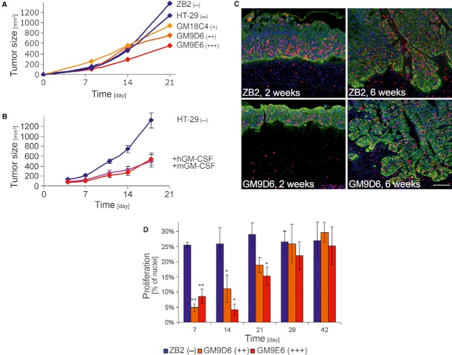
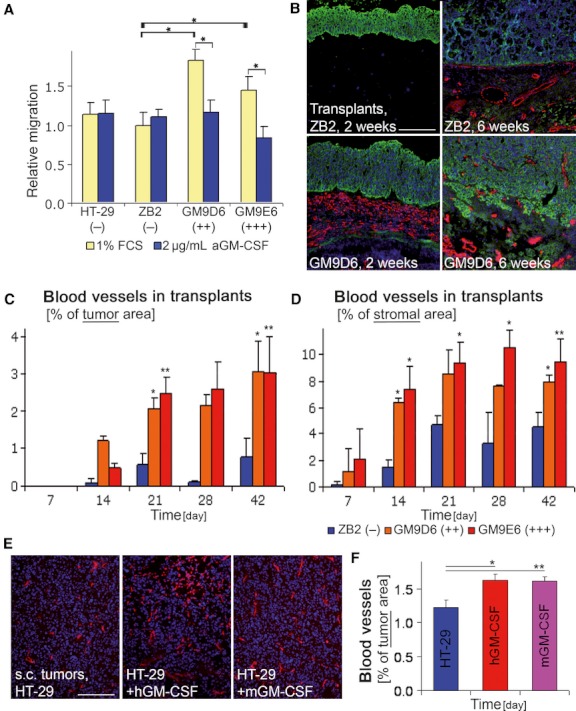
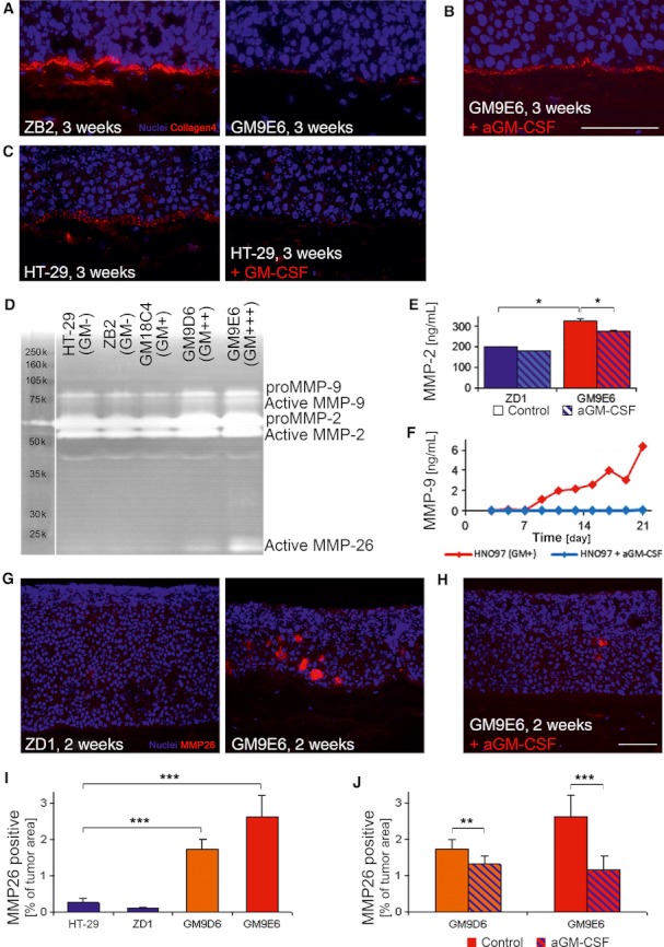
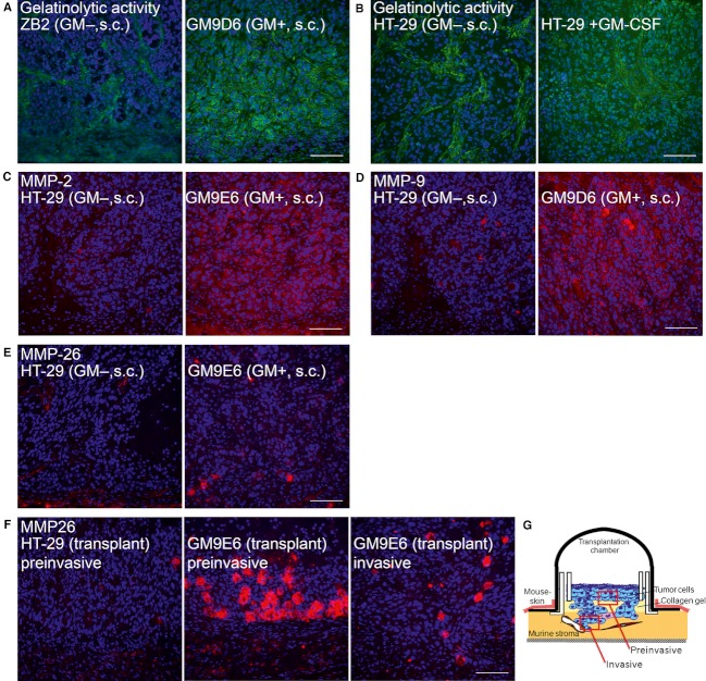
Granulocyte-macrophage colony-stimulating factor (GM-CSF) promotes tumor progression in different tumor models in an autocrine and paracrine manner. However, at the same time GM-CSF is used in cancer therapies to ameliorate neutropenia. We have previously shown in GM-CSF and G-CSF expressing or negative skin or head and neck squamous cell carcinoma that GM-CSF expression is associated with a highly angiogenic and invasive tumor phenotype. To determine the functional contribution of GM-CSF to tumor invasion, we stably transfected a GM-CSF negative colon adenocarcinoma cell line HT-29 with GM-CSF or treated the same cell line with exogenous GM-CSF. While GM-CSF overexpression and treatment reduced tumor cell proliferation and tumor growth in vitro and in vivo, respectively, it contributed to tumor progression. Together with an enhanced migratory capacity in vitro, we observed a striking increase in tumor cell invasion into the surrounding tissue concomitant with the induction of an activated tumor stroma in GM-CSF overexpressing or GM-CSF treated tumors. In a complex 3D in vitro model, enhanced GM-CSF expression was associated with a discontinued basement membrane deposition that might be mediated by the increased expression and activation of MMP-2, -9, and -26. Treatment with GM-CSF blocking antibodies reversed this effect. The increased presence and activity of these tumor cell derived proteases was confirmed in vivo. Here, expression of MMP-26 protein was predominantly located in pre- and early-invasive areas suggesting MMP-26 expression as an early event in promoting GM-CSF dependent tumor invasion.

Keywords: Colon carcinoma; GM-CSF; MMP-2; MMP-26; MMP-9; invasion.


Figures

Figure 1
Figure 1
Effect of GM-CSF on tumor growth and tumor cell proliferation in vivo. (A) While tumors of GM-CSF negative cell lines (ZB2 and HT-29) reached a size of about 1200 mm3 after 3 weeks, injection tumors of the low GM-CSF overexpressing clone (GM18C4) reached about 900 mm3, and of the high GM-CSF overexpressing clones (GM9D6 and GM9E6) about 750 and 550 mm3, respectively. (B) And while the GM-CSF negative cell line HT-29 reached a size of 1200 mm3, treatment with either hGM-CSF or mGM-CSF resulted in reduced tumor growth and a final tumor volume of about 550 mm3 after 18 days. (C) IF staining for keratin (green), proliferation (BrdU after incorporation, red), and nuclei (blue) of nude mouse surface transplant cryosections showed a reduced number of proliferating cells in 2 weeks old surface transplants of GM-CSF overexpressing GM9D6 compared with GM-CSF negative ZB2 cells, whereas no differences were detectable after 6 weeks. (D) Quantification of tumor cell proliferation in surface transplants confirmed this, showing a constantly high proliferation rate of GM-CSF negative ZB2 transplants, compared with a significantly about 75% reduced proliferations rate at day 7 in GM-CSF overexpressing transplants, increasing for 21 days to resulting in a proliferation rate comparable to transplants of GM-CSF negative cells after 28 days. Scale bar: 100 μm; *P < 0.05; **P < 0.01; d, days.
Figure 2
Figure 2
Effects of GM-CSF on tumor cell migration, invasion, and angiogenesis. (A) In a 2D scratch assay in vitro, GM-CSF overexpression (GM9D6, GM9E6) significantly enhanced tumor cell migration compared with GM-CSF negative cells (HT-29, ZB2; yellow bars), and GM-CSF blocking antibodies abrogated migration in all GM-CSF overexpressing cell lines significantly, but had no effect on GM-CSF negative cells (blue bars). (B) Immunofluorescent staining for keratin (green), CD31 positive blood vessels (red), and nuclei (blue) of cryosections from surface transplants (2 and 6 weeks). In GM-CSF negative ZB2 transplants, blood vessels rarely penetrated the collagen gel after 2 weeks and vessels reached up to the tumor border after 6 weeks with few vessels within the tumor mass. Transplants of GM-CSF overexpressing GM9E6 cells showed more early angiogenesis with extensive tumor invasion. (C) Quantification of blood vessels in the tumor area of surface transplants revealed an earlier and significantly elevated blood vessel number of GM-CSF overexpressing cells (GM9D6, GM9E6) compared with those of GM-CSF negative cells (ZB2). (D) Quantification of blood vessels in the stromal area of surface transplants also showed a significantly elevated blood vessel number of GM-CSF overexpressing cells (GM9D6, GM9E6) compared with those of GM-CSF negative cells (ZB2). (E) Immunofluorescent staining for CD31 positive blood vessels (red) and nuclei (blue) of cryosections from subcutaneous injection tumors and quantification (F). Blood vessels numbers are elevated in hGM-CSF and mGM-CSF treated tumors compared with the untreated control. Scale bar: 200 μm; *P < 0.05; **P < 0.01; d, days.
Figure 3
Figure 3
Tumor cell invasion and MMP expression in a 3D in vitro model. (A–C) Immunofluorescent staining for collagen-4 (red) and nuclei (blue) of 3D in vitro OTC cryosections (3 weeks). (A) The collagen-4 deposition at the tumor-stroma boarder decreased with increasing GM-CSF expression in the 3D in vitro OTC model. (B) GM-CSF blocking antibodies restored this collagen-4 deposition. (C) GM-CSF treatment also resulted in a decreased deposition of collagen-4 at the tumor-stroma boarder. (D) Gelatin zymographies of OTC conditioned medium revealed at day 14 an increase in pro- and active MMP-2 and MMP-9 as well as a doubled band from about 18–22 kDa that is most likely active MMP-26. (E) Significantly elevated MMP-2 secretion was verified via protein ELISA of OTC conditioned medium, as shown for GM-CSF negative (ZD1) and GM-CSF overexpressing (GM9E6) cells (unicolored bars). Moreover, GM-CSF blocking antibodies significantly inhibited MMP-2 secretion of GM-CSF overexpressing (GM9E6) but not of GM-CSF negative (ZD1) cells (striped bars). (F) For the high GM-CSF expressing HNSCC cell line HNO97, blockade with GM-CSF neutralizing antibodies resulted in complete abrogation of MMP-9, as measured by ELISA. (G) Immunofluorescent staining for MMP-26 (red) and nuclei (blue) of 3D in vitro OTC cryosections showed a strong increased cellular MMP-26 staining of GM-CSF overexpressing (GM9E6) compared with GM-CSF negative (ZD1) cells at day 14. (H) GM-CSF blocking antibodies almost abrogated MMP-26 signals of 3D in vitro OTC sections of GM-CSF overexpressing cells (GM9E6). (I) Quantification of MMP-26 staining revealed a significant increase with GM-CSF overexpression (GM-CSF negative: HT-29, ZD1; GM-CSF overexpressing: GM9D6, GM9E6). (J) GM-CSF blocking antibodies reduced this MMP-26 staining significantly in GM-CSF overexpressing clones (GM9D6, GM9E6). Scale bar: 100 μm; *P < 0.05; **P < 0.01; ***P < 0.005; d, days.
Figure 4
Figure 4
Proteolytic activity and MMP expression in vivo. (A–B) Gelatinolytic activity, as measured by in situ zymography of subcutaneous tumor cryosections, in tumors of GM-CSF negative cells (ZB2, HT-29) is located in the tumor stroma. (A) In tumors of GM-CSF overexpressing cells (GM9D6), gelatinolytic activity was also observed in the stroma, but we additionally detected a signal in the tumor cells, exceeding the stromal signal. (B) GM-CSF treatment also resulted in increased signal intensity in tumor cells. (C) Immunofluorescent (IF) staining for MMP-2 (red) and nuclei (blue) of subcutaneous tumor cryosections showed an increased staining of tumor cells in GM-CSF overexpressing (GM9E6) tumors compared with those of GM-CSF negative cells (HT-29). (D) IF staining for MMP-9 (red) and nuclei (blue) of subcutaneous tumor cryosections show, besides cellular signals in the stromal compartment, an increased staining of tumor cells in GM-CSF overexpressing (GM9D6) subcutaneous tumors compared with those of GM-CSF negative cells (HT-29). (E) IF staining of subcutaneous tumor cryosections for MMP-26 (red) and nuclei (blue) show an increased number of MMP-26 positive single tumor cells together with an increased staining of the tumor islands in GM-CSF overexpressing (GM9E6) subcutaneous tumors compared with those of GM-CSF negative cells (HT-29). (F) Moreover, IF staining of surface transplant cryosections for MMP-26 from week 4 showed a strong increased number of MMP-26 positive cells in preinvasive areas of GM-CSF overexpressing (GM9E6) compared with GM-CSF negative (HT-29) transplants. In invasive areas of the same transplants, the number of MMP-26 positive cells was downregulated again, but still exceeded the number of GM-CSF negative HT-29 transplants. (G) Schema to define the localization of “preinvasive” and “invasive” areas in surface transplants. All IF pictures depicted with the same staining are acquired under equal conditions (light intensity, exposure time, merging). Scale bar: 100 μm.

References

    1. Barreda DR, Hanington PC, Belosevic M. Regulation of myeloid development and function by colony stimulating factors. Dev. Comp. Immunol. 2004;28:509–554. - PubMed
    1. Metcalf D. The colony-stimulating factors and cancer. Nat. Rev. Cancer. 2010;10:425–434. - PMC - PubMed
    1. Williams ME, Quesenberry PJ. Hematopoietic growth factors. Hematol. Pathol. 1992;6:105–124. - PubMed
    1. Bussolino F, Colotta F, Bocchietto E, Guglielmetti A, Mantovani A. Recent developments in the cell biology of granulocyte-macrophage colony-stimulating factor and granulocyte colony-stimulating factor: activities on endothelial cells. Int. J. Clin. Lab. Res. 1993;23:8–12. - PubMed
    1. Mann A, Breuhahn K, Schirmacher P, Blessing M. Keratinocyte-derived granulocyte-macrophage colony stimulating factor accelerates wound healing: stimulation of keratinocyte proliferation, granulation tissue formation, and vascularization. J. Invest. Dermatol. 2001;117:1382–1390. - PubMed

Publication types

MeSH terms

Substances

LinkOut - more resources