. 2013 May 1:13:96.
doi: 10.1186/1471-2148-13-96.

Origin, evolution and classification of type-3 copper proteins: lineage-specific gene expansions and losses across the Metazoa

Affiliations

Origin, evolution and classification of type-3 copper proteins: lineage-specific gene expansions and losses across the Metazoa

Felipe Aguilera et al. BMC Evol Biol. .

Abstract

Background: Tyrosinases, tyrosinase-related proteins, catechol oxidases and hemocyanins comprise the type-3 copper protein family and are involved in a variety of biological processes, including pigment formation, innate immunity and oxygen transport. Although this family is present in the three domains of life, its origin and early evolution are not well understood. Previous analyses of type-3 copper proteins largely have focussed on specific animal and plant phyla.

Results: Here, we combine genomic, phylogenetic and structural analyses to show that the original type-3 copper protein possessed a signal peptide and may have been secreted (we designate proteins of this type the α subclass). This ancestral type-3 copper protein gene underwent two duplication events, the first prior to the divergence of the unikont eukaryotic lineages and the second before the diversification of animals. The former duplication gave rise to a cytosolic form (β) and the latter to a membrane-bound form (γ). Structural comparisons reveal that the active site of α and γ forms are covered by aliphatic amino acids, and the β form has a highly conserved aromatic residue in this position. The subsequent evolution of this gene family in modern lineages of multicellular eukaryotes is typified by the loss of one or more of these three subclasses and the lineage-specific expansion of one or both of the remaining subclasses.

Conclusions: The diversity of type-3 copper proteins in animals and other eukaryotes is consistent with two ancient gene duplication events leading to α, β and γ subclasses, followed by the differential loss and expansion of one or more of these subclasses in specific kingdoms and phyla. This has led to many lineage-specific type-3 copper protein repertoires and in some cases the independent evolution of functionally-classified tyrosinases and hemocyanins. For example, the oxygen-carrying hemocyanins in arthropods evolved from a β-subclass tyrosinase, whilst hemocyanins in molluscs and urochordates evolved independently from an α-subclass tyrosinase. Minor conformational changes at the active site of α, β and γ forms can produce type-3 copper proteins with capacities to either carry oxygen (hemocyanins), oxidize diphenols (catechol oxidase) or o-hydroxylate monophenols (tyrosinases) and appear to underlie some functional convergences.


Figures
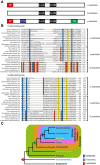
Figure 1
Figure 1
Domain architecture, copper-binding site alignment and origin of type-3 copper protein subclasses. A. Schematic representation of domain architecture of each type-3 copper protein subclass. SP: signal peptide; CYS: cysteine-rich regions; Cu(A) and Cu(B): copper-binding sites; TM: transmembrane region. B. Sequence alignment of both copper-binding sites from representatives of each type-3 copper protein subclass. Active-site histidine residues, which are important in copper-binding site conformation (blue), and conserved amino acids across all subclasses (yellow), as well as conserved amino acids restricted to specific subclasses (orange) that might have important role in structural conformation. See Additional file 1 for protein nomenclature. C. A simplified phylogenetic tree of the three domains of life showing the emergence of α-, β- and γ-subclasses.
Figure 2
Figure 2
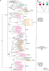
Phylogenetic analysis of the type-3 copper subclass proteins. A. A representative phylogenetic tree based on Bayesian Inference (BI), which is midpoint rooted. Statistical support is indicated at the nodes; first number, BI posterior probabilities; second number, ML bootstrap support; third number, NJ bootstrap support. Only statistical support values >50% are shown. Accession numbers of the proteins used in this tree can be found in Additional file 1. See Additional files 2, 3, 4 for detailed phylogenetic analyses of each type-3 copper protein subclass. B. Phylogenetic relationship and functionalities found in type-3 copper subclasses. T, tyrosinase; C, catechol oxidases; H, hemocyanin. Species were labelled according to a specific colour code as follows: black: Eubacteria; sky blue: Archaeobacteria; purple: Plantae; orange: Chromoalveaolata; dark blue: Amoebozoa; dark green: Fungi; yellow: Porifiera; light green: Cnidaria; magenta: Protostomia (Mollusca, Annelida, Platyhelminthes, Nematoda, Arthropoda and Onycophora); brown: Deuterostomia (Hemichordata, Cephalochordata, Urochordata and Vertebrata).
Figure 3
Figure 3
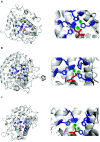
Binuclear active site of tyrosinase proteins and placeholder residue blocking the entrance of the substrate. Stereo view of tyrosinase active site region with both Cu-binding sites is presented. Copper ions are depicted in green, Cu(A) is shown on the left and Cu(B) is shown on the right. The yellow sphere represents a dioxygen molecule. In each structure, the occupation and positioning of copper varies. The six copper-coordinating histidine ligands coordinating the structural conformation of the active site are shown in dark blue. In addition, the placeholder residue that reaches into the active site above Cu(A) and blocks the substrate-binding pocket and the entrance of the substrate into the active site is indicated in red. Differences in the orientation and size of the blocking residue are key to the enzymatic activity of tyrosinases. Representatives of three tyrosinase proteins from α- subclass are shown. A. Mollusc tyrosinase, B. Nematode tyrosinase and C. Cnidarian tyrosinase.
Figure 4
Figure 4
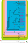
Summary of gene expansions and losses of type-3 copper protein subclasses. The phylogenetic relationships between the species under study are shown. The ancestral form (α-subclass, red colour code) arose early in the evolution of life. The β-subclass (green colour code) emerged before the divergence of unikont lineages. Finally, a γ-subclass (blue colour code) emerged as a second duplication of the α-subclass ancestor prior to metazoan diversification. Coloured crosses denote cases for which losses of particular subclasses have occurred in specific lineages. Gene expansions are indicated by coloured dots for each lineage. Species are labelled according to colour code shown in Figure 2.

Similar articles

Cited by

References

    1. Solomon EI, Baldwin MJ, Lowery MD. Electronic structures of active sites in copper proteins: contributions to reactivity. Chem Rev. 1992;92:521–542. doi: 10.1021/cr00012a003. - DOI
    1. Solomon EI, Sundaram UM, Machonkin TE. Multicopper oxidases and oxygenases. Chem Rev. 1996;96:2563–2605. doi: 10.1021/cr950046o. - DOI - PubMed
    1. Festa RA, Thiele DJ. Copper: An essential metal in biology. Curr Biol. 2011;21(21):R877–R883. doi: 10.1016/j.cub.2011.09.040. - DOI - PMC - PubMed
    1. Andreini C, Banci L, Bertini I, Rosato A. Occurrence of copper proteins through the three domains of life: A bioinformatic approach. Proteome Res. 2008;7:209–216. doi: 10.1021/pr070480u. - DOI - PubMed
    1. van Gelder CW, Flurkey WH, Wichers HJ. Sequence and structural features of plant and fungal tyrosinases. Phytochemistry. 1997;45(7):1309–1323. doi: 10.1016/S0031-9422(97)00186-6. - DOI - PubMed

Publication types

MeSH terms

LinkOut - more resources