

A virtual trial framework for quantifying the detectability of masses in breast tomosynthesis projection data
- PMID: 23635284
- PMCID: PMC3651214
- DOI: 10.1118/1.4800501
A virtual trial framework for quantifying the detectability of masses in breast tomosynthesis projection data
Abstract
Purpose: Digital breast tomosynthesis (DBT) is a promising breast cancer screening tool that has already begun making inroads into clinical practice. However, there is ongoing debate over how to quantitatively evaluate and optimize these systems, because different definitions of image quality can lead to different optimal design strategies. Powerful and accurate tools are desired to extend our understanding of DBT system optimization and validate published design principles.
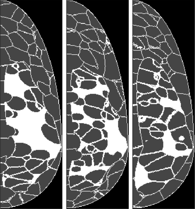
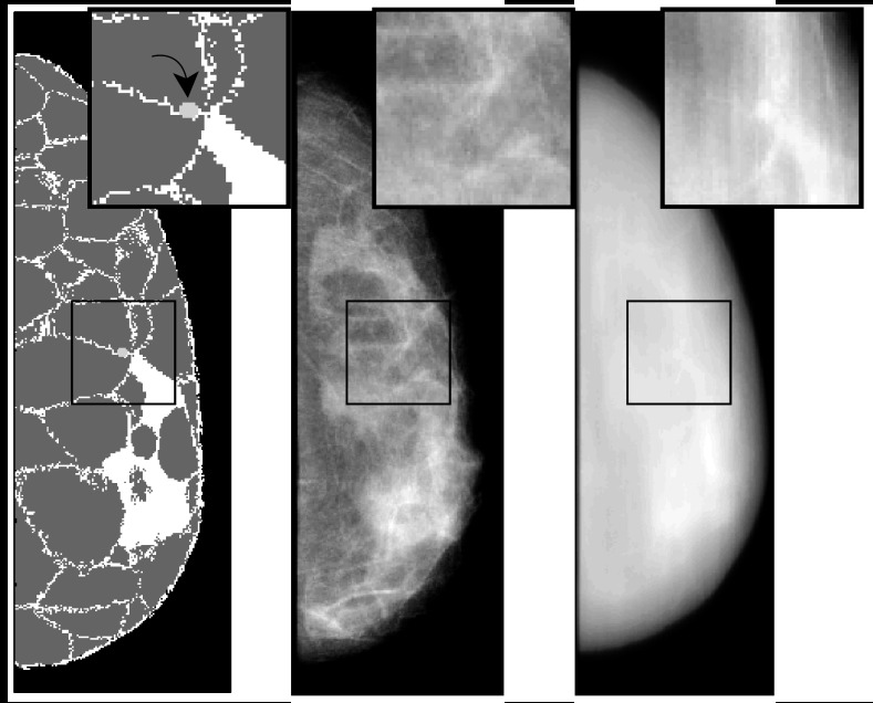
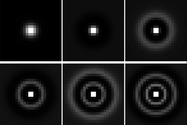
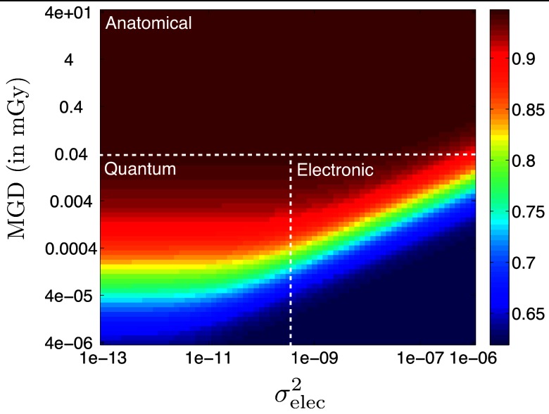
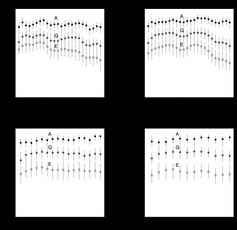
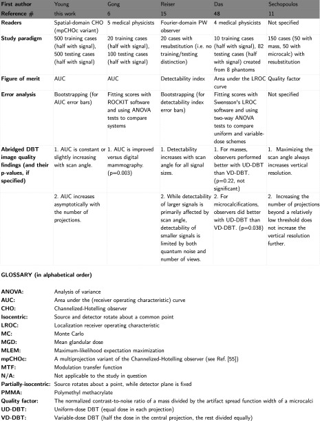
Methods: The authors developed a virtual trial framework for task-specific DBT assessment that uses digital phantoms, open-source x-ray transport codes, and a projection-space, spatial-domain observer model for quantitative system evaluation. The authors considered evaluation of reconstruction algorithms as a separate problem and focused on the information content in the raw, unfiltered projection images. Specifically, the authors investigated the effects of scan angle and number of angular projections on detectability of a small (3 mm diameter) signal embedded in randomly-varying anatomical backgrounds. Detectability was measured by the area under the receiver-operating characteristic curve (AUC). Experiments were repeated for three test cases where the detectability-limiting factor was anatomical variability, quantum noise, or electronic noise. The authors also juxtaposed the virtual trial framework with other published studies to illustrate its advantages and disadvantages.
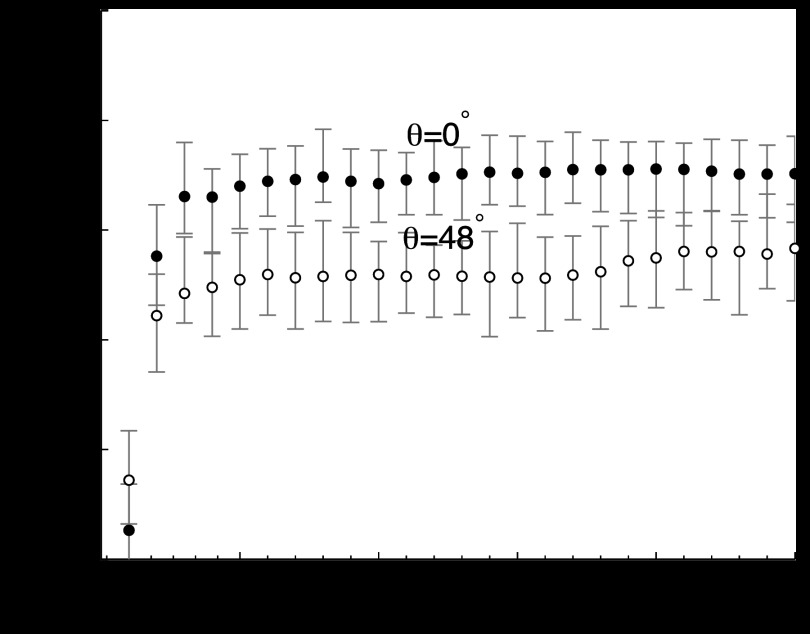
Results: The large number of variables in a virtual DBT study make it difficult to directly compare different authors' results, so each result must be interpreted within the context of the specific virtual trial framework. The following results apply to 25% density phantoms with 5.15 cm compressed thickness and 500 μm(3) voxels (larger 500 μm(2) detector pixels were used to avoid voxel-edge artifacts): 1. For raw, unfiltered projection images in the anatomical-variability-limited regime, AUC appeared to remain constant or increase slightly with scan angle. 2. In the same regime, when the authors fixed the scan angle, AUC increased asymptotically with the number of projections. The threshold number of projections for asymptotic AUC performance depended on the scan angle. In the quantum- and electronic-noise dominant regimes, AUC behaviors as a function of scan angle and number of projections sometimes differed from the anatomy-limited regime. For example, with a fixed scan angle, AUC generally decreased with the number of projections in the electronic-noise dominant regime. These results are intended to demonstrate the capabilities of the virtual trial framework, not to be used as optimization rules for DBT.
Conclusions: The authors have demonstrated a novel simulation framework and tools for evaluating DBT systems in an objective, task-specific manner. This framework facilitates further investigation of image quality tradeoffs in DBT.
Figures


References
-
- Fahrig R., Maidment A. D. A., and Yaffe M. J., “Optimization of peak kilovoltage and spectral shape for digital mammography,” Proc. SPIE 1651, 74–83 (1992). 10.1117/12.59405 - DOI
Publication types
MeSH terms
Grants and funding
LinkOut - more resources
Full Text Sources
Other Literature Sources
Medical
