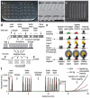
Multiplexed electrochemical protein detection and translation to personalized cancer diagnostics
- PMID: 23635325
- PMCID: PMC3674208
- DOI: 10.1021/ac401058v
Multiplexed electrochemical protein detection and translation to personalized cancer diagnostics
Abstract
Measuring diagnostic panels of multiple proteins promises a new, personalized approach to early detection and therapy of diseases like cancer. Levels of biomarker proteins in patient serum can provide a continually updated record of disease status. Research in electrochemical detection of proteins has produced exquisitely sensitive approaches. Most utilize ELISA-like sandwich immunoassays incorporating various aspects of nanotechnology. Several of these ultrasensitive methodologies have been extended to microfluidic multiplexed protein detection, but engineered solutions are needed to measure more proteins in a single device from a small patient sample such as a drop of blood or tissue lysate. To achieve clinical or point-of-care (POC) use, simplicity and low cost are essential. In multiplexed microfluidic immunoassays, required reagent additions and washing steps pose a significant problem calling for creative engineering. A grand challenge is to develop a general cancer screening device to accurately measure 50-100 proteins in a simple, cost-effective fashion. This will require creative solutions to simplified reagent addition and multiplexing.
Figures


Similar articles
-
Multiplex Immunosensor Arrays for Electrochemical Detection of Cancer Biomarker Proteins.Electroanalysis. 2016 Nov;28(11):2644-2658. doi: 10.1002/elan.201600183. Epub 2016 Jun 7. Electroanalysis. 2016. PMID: 28592919 Free PMC article.
-
Printed Electrodes in Microfluidic Arrays for Cancer Biomarker Protein Detection.Biosensors (Basel). 2020 Sep 7;10(9):115. doi: 10.3390/bios10090115. Biosensors (Basel). 2020. PMID: 32906644 Free PMC article. Review.
-
Toward Personalized Cancer Treatment: From Diagnostics to Therapy Monitoring in Miniaturized Electrohydrodynamic Systems.Acc Chem Res. 2019 Aug 20;52(8):2113-2123. doi: 10.1021/acs.accounts.9b00192. Epub 2019 Jul 11. Acc Chem Res. 2019. PMID: 31293158 Review.
-
Ultrasensitive detection of cancer biomarkers in the clinic by use of a nanostructured microfluidic array.Anal Chem. 2012 Jul 17;84(14):6249-55. doi: 10.1021/ac301392g. Epub 2012 Jul 3. Anal Chem. 2012. PMID: 22697359 Free PMC article.
-
Enabling Multiplexed Electrochemical Detection of Biomarkers with High Sensitivity in Complex Biological Samples.Acc Chem Res. 2021 Sep 21;54(18):3529-3539. doi: 10.1021/acs.accounts.1c00382. Epub 2021 Sep 3. Acc Chem Res. 2021. PMID: 34478255
Cited by
-
Microfluidic integration of regeneratable electrochemical affinity-based biosensors for continual monitoring of organ-on-a-chip devices.Nat Protoc. 2021 May;16(5):2564-2593. doi: 10.1038/s41596-021-00511-7. Epub 2021 Apr 28. Nat Protoc. 2021. PMID: 33911259
-
Multiplexed Electrochemical Immunosensors for Clinical Biomarkers.Sensors (Basel). 2017 Apr 27;17(5):965. doi: 10.3390/s17050965. Sensors (Basel). 2017. PMID: 28448466 Free PMC article. Review.
-
Glucose biosensor based on open-source wireless microfluidic potentiostat.Sens Actuators B Chem. 2019 Jul 1;290:616-624. doi: 10.1016/j.snb.2019.02.031. Epub 2019 Feb 17. Sens Actuators B Chem. 2019. PMID: 32395016 Free PMC article.
-
Multiplex Immunosensor Arrays for Electrochemical Detection of Cancer Biomarker Proteins.Electroanalysis. 2016 Nov;28(11):2644-2658. doi: 10.1002/elan.201600183. Epub 2016 Jun 7. Electroanalysis. 2016. PMID: 28592919 Free PMC article.
-
An electrochemical clamp assay for direct, rapid analysis of circulating nucleic acids in serum.Nat Chem. 2015 Jul;7(7):569-75. doi: 10.1038/nchem.2270. Epub 2015 Jun 1. Nat Chem. 2015. PMID: 26100805
References
-
- Atkinson AJ, Colburn WA, DeGruttola VG, et al. Clin. Pharmacol. Ther. 2001;69:89–95. - PubMed
-
- Manne U, Srivastava RG, Srivastava S. Drug. Discov. Today. 2005;10:965–976. - PubMed
-
- Ludwig JA, Weinstein JN. Nature Rev. Cancer. 2005;5:845–856. - PubMed
-
- Kulasingam V, Diamandis EP. Nature Clin. Pract. Oncol. 2008;5:588–599. - PubMed
Publication types
MeSH terms
Substances
Grants and funding
LinkOut - more resources
Full Text Sources
Other Literature Sources

