. 2013 May 1:11:106.
doi: 10.1186/1479-5876-11-106.

Treatment of malignant effusion by oncolytic virotherapy in an experimental subcutaneous xenograft model of lung cancer

Affiliations

Treatment of malignant effusion by oncolytic virotherapy in an experimental subcutaneous xenograft model of lung cancer

Stephanie Weibel et al. J Transl Med. .

Abstract

Background: Malignant pleural effusion (MPE) is associated with advanced stages of lung cancer and is mainly dependent on invasion of the pleura and expression of vascular endothelial growth factor (VEGF) by cancer cells. As MPE indicates an incurable disease with limited palliative treatment options and poor outcome, there is an urgent need for new and efficient treatment options.

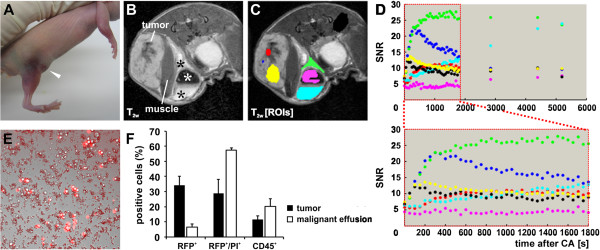
Methods: In this study, we used subcutaneously generated PC14PE6 lung adenocarcinoma xenografts in athymic mice that developed subcutaneous malignant effusions (ME) which mimic pleural effusions of the orthotopic model. Using this approach monitoring of therapeutic intervention was facilitated by direct observation of subcutaneous ME formation without the need of sacrificing mice or special imaging equipment as in case of MPE. Further, we tested oncolytic virotherapy using Vaccinia virus as a novel treatment modality against ME in this subcutaneous PC14PE6 xenograft model of advanced lung adenocarcinoma.

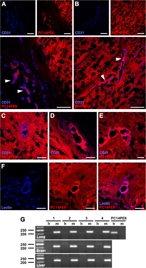
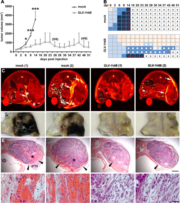
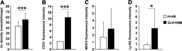
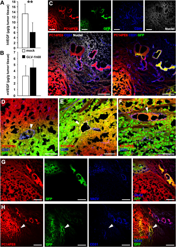
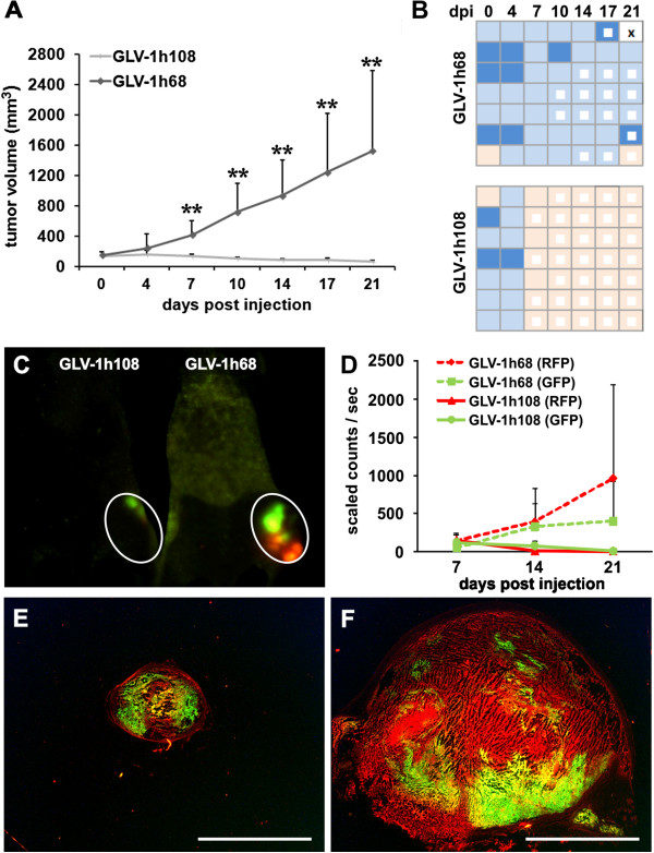
Results: We demonstrated significant therapeutic efficacy of Vaccinia virus treatment of both advanced lung adenocarcinoma and tumor-associated ME. We attribute the efficacy to the virus-mediated reduction of tumor cell-derived VEGF levels in tumors, decreased invasion of tumor cells into the peritumoral tissue, and to viral infection of the blood vessel-invading tumor cells. Moreover, we showed that the use of oncolytic Vaccinia virus encoding for a single-chain antibody (scAb) against VEGF (GLAF-1) significantly enhanced mono-therapy of oncolytic treatment.

Conclusions: Here, we demonstrate for the first time that oncolytic virotherapy using tumor-specific Vaccinia virus represents a novel and promising treatment modality for therapy of ME associated with advanced lung cancer.


Figures

Figure 1
Figure 1
Generation of subcutaneous PC14PE6-RFP lung adenocarcinomas and tumor-associated ME. (A) Photographic image of a mouse bearing a subcutaneous PC14PE6-RFP tumor on the right flank 14 dpim showing a hematoma around the tumor site and the beginning of exudate accumulation in the groin (arrowhead). (B-C) exemplary T1w MRI of a PC14PE6-RFP-bearing mouse 22 dpim revealing tumor-associated effusion in the groin containing fluid (black asterisks, B) and solid/semi-solid cellular material (white asterisk, B). Using T1w MRI the time-dependent distribution of (iv) injected Gd-DTPA was recorded in different tumor regions and areas of the ME as indicated by regions of interest (ROIs) in (C); the corresponding SNR over time of the different ROIs were plotted in (D). ROI coloring: red – necrotic tumor region, blue – enlarged blood vessel, yellow – non-necrotic tumor region, green – outer effusion region, pink – solid effusion content, light blue – fluid effusion content, black – control muscle tissue. (E) micro-photographic image overlayed with the red fluorescent image of an exudate smear revealing numerous erythrocytes as well as red-fluorescent tumor cells and non-fluorescent cells. (F) FACS analysis of PC14PE6-RFP tumors (28 dpim, n = 4) and ME (n = 3); shown are the percentage of RFP-positive tumor cells, propidium iodide (PI)/RFP-positive dead tumor cells, and CD45-positive immune cells in tumors and ME.
Figure 2
Figure 2
Enlarged tumor cell-containing CD31-positive blood vessels (TCCBVs) in PC14PE6-RFP tumors. (A-F) Confocal microscopic images of 15 μm-thick PC14PE6-RFP tumor sections 21 dpim showed enlarged CD31-positive blood vessels (blue) containing RFP-expressing tumor cells (red, arrowheads) in peritumoral regions (A) and tumor border areas (B). In tumor sections enlarged CD31-positive TCCBVs appear in various morphological formations resembling glomeruloid bodies (C) or garland-like vessels with intussusceptive vascular growth (D). Functionality of enlarged TCCBVs was confirmed by co-localization of CD41-positive platelets with garland-like TCCBVs (E) and Lectin-labeling of TCCBVs upon systemic injection of Lectin into PC14PE6-RFP-bearing mice. Both microscopic images indicate the connection of TCCBVs to the blood circulation. (G) RT-PCR analysis of lung, brain and liver homogenates of end-stage PC14PE6-RFP-bearing mice (28 dpim) using human- (h, 205 bp) and mouse-specific (m, 216 bp) ß-actin primers (n = 4). Human PC14PE6-RFP cells were used as a positive control for human ß-actin and as a negative control for mouse ß-actin. All images are representative examples. Scale bars represent 300 μm (A, B, F), 75 μm (C, D), and 40 μm (E).
Figure 3
Figure 3
Therapy of ME in PC14PE6-RFP tumor-bearing mice upon systemic injection of GLV-1h68. (A-B) PC14PE6-RFP tumor-bearing mice were either mock-infected or treated with 1 × 107 pfu GLV-1h68 (iv) 14 dpim (n = 6). (A) PC14PE6-RFP tumor growth was monitored by measuring the tumor volume revealing a continuous tumor growth in the mock-infected group and a significantly retarded growth rate in the GLV-1h68-treated group; all mice of the mock-infected group were euthanized due to tumor burden and ME formation 14 dpi; in the GLV-1h68-treated group two mice were euthanized due to ME formation (23, 42 dpi) before end of the study. Shown are the mean values +/− standard deviations. (B) same mice as shown in A were individually monitored for ME formation during the study; ME formation was evaluated by skin color of the tumor area (hematoma) and exudate accumulation in the groin; skin color index indicating increased hematoma: skin-coloured – light blue – middle blue – dark blue; red dot: palpable cyst in the groin (exudate accumulation), white square: area of tumor necrosis, cross: euthanization. (C) in vivo MR T2 maps of two mock- and two GLV-1h68-infected PC14PE6-RFP tumor-bearing mice 7 dpi. Tumor-associated effusions (arrow) with accumulation of solid/semi-solid cellular components (asterisk) are indicated in T2 images. The T2 maps are scaled from 0 to 200 ms. (D) the corresponding photographic images of the paraformaldehyde-fixed and decalcified mouse abdomen demonstrated the extended hematoma in the peritumoral area of mock-infected mice; in GLV-1h68-treated mice no peritumoral hematoma was detected. (E, F) H&E staining of the corresponding whole abdominal mouse sections revealed erythrocytes-containing blood lakes (black asterisks, E) in mock-treated and necrotic tumor areas (white asterisk, E) in GLV-1h68-treated mice. Different histopathology of the tumor margin of mock- and GLV-1h68-treated tumors (arrowheads, E) is shown with higher magnification in (F). All images are representative examples. Scale bars represent 2 mm (E) 20 μm (F).
Figure 4
Figure 4
VACV infection-induced activation of the tumor vasculature. (A-D) PC14PE6-RFP tumor-bearing mice were either mock-infected or treated with 1 × 107 pfu GLV-1h68 (iv) 14 dpim (n = 3). Seven dpi tumors were harvested and histological tissue sections were performed. The blood vessel density (A) was determined by counting the vessels/image in both groups and the mean fluorescence intensity of endothelial-associated CD31 was shown in (B). The content of either antigen-presenting MHCII-positive cells (monocytes, macrophages, dendritic cells, and B cells) (C) or Ly-6G-positive neutrophils (D) in mock- and GLV-1h68-treated tumors was determined by quantification of the mean fluorescence intensity of the antibody-labeled tumor sections.
Figure 5
Figure 5
VACV treatment decreased tumor cell-produced hVEGF and infected TCCBVs in PC14PE6-RFP tumors. (A, B) PC14PE6-RFP tumor-bearing mice were either mock-infected or treated with 1 × 107 pfu GLV-1h68 (iv) 14 dpim. Tumor homogenates of mock- and GLV-1h68-treated tumors (7dpi, n = 6) were used for ELISA of human VEGF (A) and murine VEGF (B). Shown are the mean values +/− standard deviations. (C-F) Histological analysis of GLV-1h68-infected PC14PE6-RFP tumor sections 7 dpi revealed a GLV-1h68-infection of TCCBVs; PC14PE6-RFP tumor cells (red), GLV-1h68-infected cells express GFP (green), blood vessels were labeled with the CD31 antibody (blue), nuclei were labeled with Hoechst 33324 (white). All the different morphological formations of TCCBVs such as glomeruloid bodies (D) and garland-like vessels (E, F) were GFP-positive. (G) anti-VACV antibody labeling (blue) of tumor sections revealed VACV-positive labeling in GFP-positive areas. (H) Histological analysis of GLV-1h68-infected PC14PE6-RFP tumor sections revealed GLV-1h68-infection of invasive PC14PE6-RFP tumor cells at the tumor margin (arrowhead); PC14PE6-RFP tumor cells (red), GLV-1h68-infected cells (green), CD31-positive blood vessels (blue). All images are representative examples. Scale bars represent 300 μm (C, G), 75 μm (D-F), and 150 μm (H).
Figure 6
Figure 6
Enhanced therapeutic efficacy in tumor xenografts with ME using an anti-VEGF scAb-encoding VACV. (A-F) PC14PE6-RFP tumor-bearing mice were either iv infected with 1 × 107 pfu GLV-1h68 or 1 x10⁷ GLV-1h108 14 dpim (n = 7). (A) PC14PE6-RFP tumor growth was monitored over 21 days pi by measuring the tumor volume revealing a continuous tumor growth in the GLV-1h68-infected group and a significant tumor regression in the GLV-1h108 treated group. Shown are the mean values +/− standard deviations. (B) same mice as shown in A were individually monitored for ME formation during the study; ME formation was evaluated by skin color of the tumor area (hematoma); skin color index indicating increased hematoma: skin-coloured – light blue – middle blue; white square: area of tumor necrosis, cross: euthanization. (C, D) real-time fluorescence imaging of tumor growth (RFP) and viral infection (GFP) during growth curve analysis at day 7, 14, and 21 pi using the Maestro EX imaging system. (C) representative fluorescence image of GLV-1h68- and GLV-1h108-infected PC14PE6-RFP-tumor-bearing mice 14 dpi. (E,F) representative microscopic images of 15 μm-thick PC14PE6-RFP tumor sections 21 dpi either infected with GLV-1h108 (E) or GLV-1h68 (F); PC14PE6-RFP tumor cells (red), viral infected GFP-expressing cells (green). All images are representative examples. Scale bars represent 5 mm (E, F).

Similar articles

Cited by

References

    1. Siegel R, Naishadham D, Jemal A. Cancer statistics, 2012. CA Canc J Clin. 2012;62:10–29. doi: 10.3322/caac.20138. - DOI - PubMed
    1. Uzbeck MH, Almeida FA, Sarkiss MG, Morice RC, Jimenez CA, Eapen GA, Kennedy MP. Management of malignant pleural effusions. Adv Ther. 2010;27:334–347. doi: 10.1007/S12325-010-0031-8. - DOI - PubMed
    1. Bennett R, Maskell N. Management of malignant pleural effusions. Curr Opin Pulm Med. 2005;11:296–300. - PubMed
    1. Lombardi G, Zustovich F, Nicoletto MO, Donach M, Artioli G, Pastorelli D. Diagnosis and treatment of malignant pleural effusion: a systematic literature review and new approaches. Am J Clin Oncol. 2010;33:420–423. doi: 10.1097/COC.0b013e3181aacbbf. - DOI - PubMed
    1. Grove CS, Lee YCG. Vascular endothelial growth factor: the key mediator in pleural effusion formation. Curr Opin Pulm Med. 2002;8:294–301. doi: 10.1097/00063198-200207000-00009. - DOI - PubMed

Publication types

Substances