

Conserved roles of ems/Emx and otd/Otx genes in olfactory and visual system development in Drosophila and mouse
- PMID: 23635521
- PMCID: PMC3866872
- DOI: 10.1098/rsob.120177
Conserved roles of ems/Emx and otd/Otx genes in olfactory and visual system development in Drosophila and mouse
Abstract
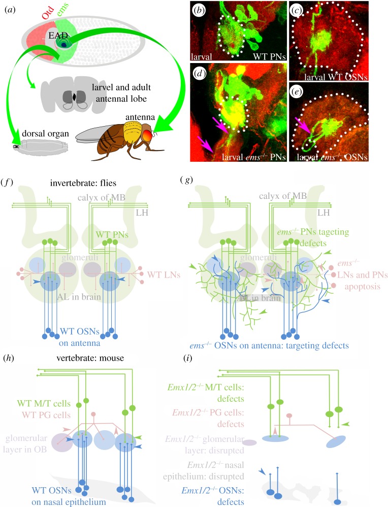
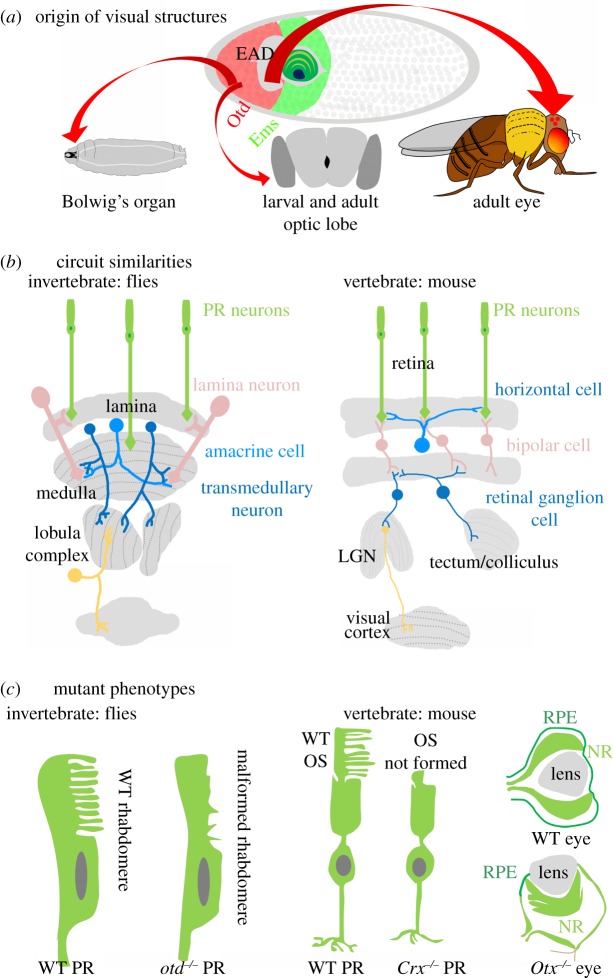
The regional specialization of brain function has been well documented in the mouse and fruitfly. The expression of regulatory factors in specific regions of the brain during development suggests that they function to establish or maintain this specialization. Here, we focus on two such factors-the Drosophila cephalic gap genes empty spiracles (ems) and orthodenticle (otd), and their vertebrate homologues Emx1/2 and Otx1/2-and review novel insight into their multiple crucial roles in the formation of complex sensory systems. While the early requirement of these genes in specification of the neuroectoderm has been discussed previously, here we consider more recent studies that elucidate the later functions of these genes in sensory system formation in vertebrates and invertebrates. These new studies show that the ems and Emx genes in both flies and mice are essential for the development of the peripheral and central neurons of their respective olfactory systems. Moreover, they demonstrate that the otd and Otx genes in both flies and mice are essential for the development of the peripheral and central neurons of their respective visual systems. Based on these recent experimental findings, we discuss the possibility that the olfactory and visual systems of flies and mice share a common evolutionary origin, in that the conserved visual and olfactory circuit elements derive from conserved domains of otd/Otx and ems/Emx action in the urbilaterian ancestor.
Keywords: Emx1/2, Otx1/2; empty spiracles; evolutionary conservation; orthodenticle; sensory systems.
Figures
Similar articles
-
Analysis of the Otd-dependent transcriptome supports the evolutionary conservation of CRX/OTX/OTD functions in flies and vertebrates.Dev Biol. 2008 Mar 15;315(2):521-34. doi: 10.1016/j.ydbio.2007.12.017. Epub 2008 Jan 31. Dev Biol. 2008. PMID: 18241855 Free PMC article.
-
Murine Otx1 and Drosophila otd genes share conserved genetic functions required in invertebrate and vertebrate brain development.Development. 1998 May;125(9):1691-702. doi: 10.1242/dev.125.9.1691. Development. 1998. PMID: 9521907
-
Otx genes in brain morphogenesis.Prog Neurobiol. 2001 May;64(1):69-95. doi: 10.1016/s0301-0082(00)00042-3. Prog Neurobiol. 2001. PMID: 11250063 Review.
-
Evolutionary conservation of otd/Otx2 transcription factor action: a genome-wide microarray analysis in Drosophila.Genome Biol. 2002;3(4):RESEARCH0015. doi: 10.1186/gb-2002-3-4-research0015. Epub 2002 Mar 14. Genome Biol. 2002. PMID: 11983056 Free PMC article.
-
Otx genes in the development and evolution of the vertebrate brain.Int J Dev Neurosci. 2001 Jul;19(4):353-63. doi: 10.1016/s0736-5748(01)00003-x. Int J Dev Neurosci. 2001. PMID: 11378295 Review.
Cited by
-
The homeodomain transcription factor Phox2 in the stellate ganglion of the squid Loligo pealei.Biol Open. 2015 Jun 26;4(8):954-60. doi: 10.1242/bio.012476. Biol Open. 2015. PMID: 26116657 Free PMC article.
-
Heterochrony in orthodenticle expression is associated with ommatidial size variation between Drosophila species.BMC Biol. 2025 Feb 4;23(1):34. doi: 10.1186/s12915-025-02136-8. BMC Biol. 2025. PMID: 39901145 Free PMC article.
-
Deciphering the Role of Emx1 in Neurogenesis: A Neuroproteomics Approach.Front Mol Neurosci. 2016 Oct 17;9:98. doi: 10.3389/fnmol.2016.00098. eCollection 2016. Front Mol Neurosci. 2016. PMID: 27799894 Free PMC article.
-
Loss of miR-210 leads to progressive retinal degeneration in Drosophila melanogaster.Life Sci Alliance. 2019 Jan 22;2(1):e201800149. doi: 10.26508/lsa.201800149. Print 2019 Feb. Life Sci Alliance. 2019. PMID: 30670478 Free PMC article.
-
The neuromuscular system of Pycnophyes kielensis (Kinorhyncha: Allomalorhagida) investigated by confocal laser scanning microscopy.Evodevo. 2016 Nov 28;7:25. doi: 10.1186/s13227-016-0062-6. eCollection 2016. Evodevo. 2016. PMID: 27933139 Free PMC article.
References
-
- Dalton D, Chadwick R, McGinnis W. 1989. Expression and embryonic function of empty spiracles: a Drosophila homeo box gene with two patterning functions on the anterior–posterior axis of the embryo. Genes Dev. 3, 1940–195610.1101/gad.3.12a.1940 (doi:10.1101/gad.3.12a.1940) - DOI - DOI - PubMed
-
- Finkelstein R, Perrimon N. 1990. The orthodenticle gene is regulated by bicoid and torso and specifies Drosophila head development. Nature 346, 485–48810.1038/346485a0 (doi:10.1038/346485a0) - DOI - DOI - PubMed
-
- Jurgens G, Hartenstein V. 1993. The terminal regions of the body pattern. In The development of Drosophila melanogaster, vol. 1 (eds Bate M, Martinez-Arias A.), pp. 687–746 Plainview, NY: Cold Spring Harbor Laboratory Press
-
- Gao Q, Finkelstein R. 1998. Targeting gene expression to the head: the Drosophila orthodenticle gene is a direct target of the Bicoid morphogen. Development 125, 4185–4193 - PubMed
Publication types
MeSH terms
Substances
LinkOut - more resources
Full Text Sources
Other Literature Sources
Molecular Biology Databases
Miscellaneous
