Review
. 2013 May 1;3(5):130023.
doi: 10.1098/rsob.130023.

Evolutionary cell biology of chromosome segregation: insights from trypanosomes

Affiliations
Review

Evolutionary cell biology of chromosome segregation: insights from trypanosomes

Bungo Akiyoshi et al. Open Biol. .

Abstract

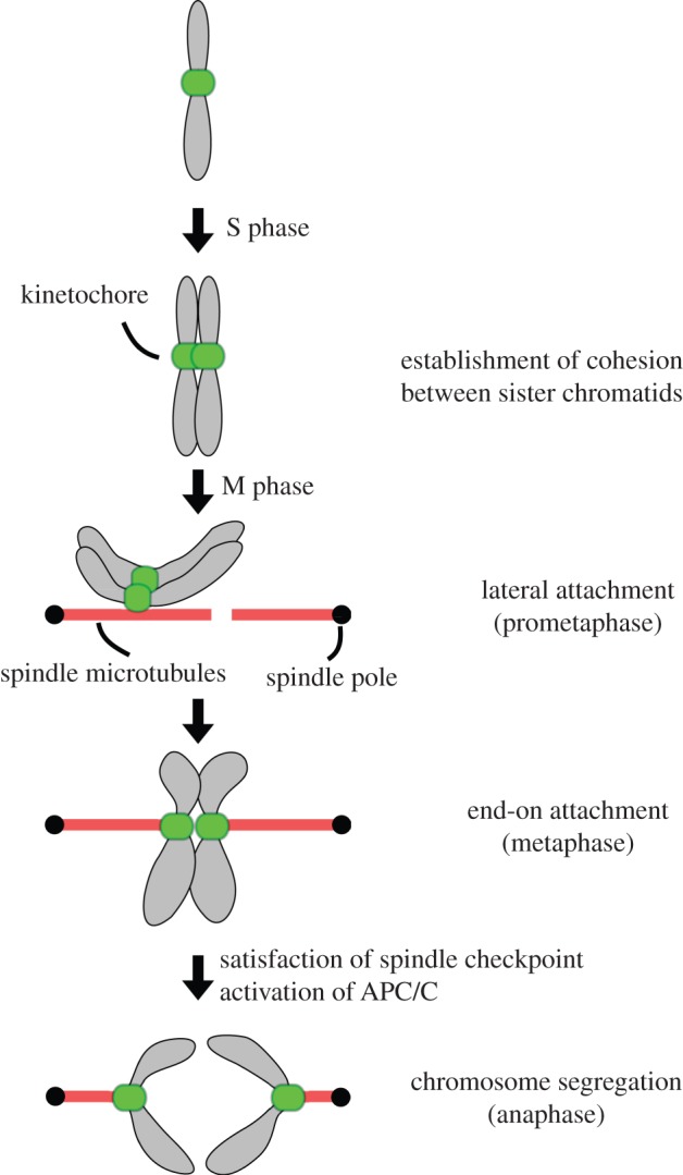
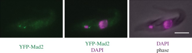
Faithful transmission of genetic material is essential for the survival of all organisms. Eukaryotic chromosome segregation is driven by the kinetochore that assembles onto centromeric DNA to capture spindle microtubules and govern the movement of chromosomes. Its molecular mechanism has been actively studied in conventional model eukaryotes, such as yeasts, worms, flies and human. However, these organisms are closely related in the evolutionary time scale and it therefore remains unclear whether all eukaryotes use a similar mechanism. The evolutionary origins of the segregation apparatus also remain enigmatic. To gain insights into these questions, it is critical to perform comparative studies. Here, we review our current understanding of the mitotic mechanism in Trypanosoma brucei, an experimentally tractable kinetoplastid parasite that branched early in eukaryotic history. No canonical kinetochore component has been identified, and the design principle of kinetochores might be fundamentally different in kinetoplastids. Furthermore, these organisms do not appear to possess a functional spindle checkpoint that monitors kinetochore-microtubule attachments. With these unique features and the long evolutionary distance from other eukaryotes, understanding the mechanism of chromosome segregation in T. brucei should reveal fundamental requirements for the eukaryotic segregation machinery, and may also provide hints about the origin and evolution of the segregation apparatus.

Keywords: CENP-A; Trypanosoma brucei; chromosome segregation; kinetochores; kinetoplastids; mitosis.


Figures

Figure 1.
Figure 1.
Mitotic chromosome segregation. Chromosomes are duplicated during S phase, and cohesion is established between sister chromatids. When cells enter mitosis, a bipolar spindle is assembled. Kinetochores initially form lateral attachments to spindle microtubules, which are then converted to end-on attachments. When all chromosomes form bi-oriented attachments (i.e. sister kinetochores attach to microtubules emanating from opposite poles), the spindle checkpoint is satisfied and the APC/C gets activated. This leads to the dissolution of cohesion so that the sister chromatids segregate away from each other.
Figure 2.
Figure 2.
Current eukaryotic phylogenetic tree. In this unrooted tree, eukaryotes are divided into six supergroups, Opisthokonta, Amoebozoa, Excavata, Archaeplastida, SAR (stramenopiles, alveolates and rhizaria) and CCTH (cryptophytes, centrohelids, telonemids and haptophytes). Representative organisms whose draft genome sequences are available are shown as examples. The tree has been redrawn and modified from [30]. Branch lengths are arbitrary.
Figure 3.
Figure 3.
Diagram of the cell division cycle in T. brucei procyclic (insect) form cells. (a) G1 cells possess a single kinetoplast and nucleus (termed 1K1N) as well as an attached flagellum. (b) As the cell cycle progresses, a new basal body forms and nucleates a new flagellum. The nucleus is still in S phase when kinetoplast DNA shows an elongated morphology. (c) Segregation of basal bodies leads to the separation of attached kinetoplast. These cells are termed 2K1N. (d) Cells enter nuclear M phase, and chromosome segregation occurs. (e) Nuclear division is complete. These cells are termed 2K2N. (f) Cleavage furrow ingression occurs between the two flagella. (g) At the end of the cell cycle, two daughter cells are formed, and each cell inherits a single kinetoplast, nucleus and flagellum.
Figure 4.
Figure 4.
Chromosome structure and organization. (a) Diagram of the three different types of chromosome in T. brucei. Essentially, all housekeeping genes are encoded in megabase chromosomes and are expressed in polycistronic transcription units. The centromere is located in a transcriptional strand-switch region of megabase chromosomes, while such a centromere appears absent from intermediate and minichromosomes. The core of minichromosomes consists of the 177 bp repeats and is constructed in a palindromic manner with a single inversion point in the centre. The complete intermediate chromosome structure is not known, but 177 bp repeats are present. (b) Diagram of the centromeric region of T. cruzi chromosome 3 and T. brucei chromosome 1, based on [–115]. Various retro-elements are found in both species (e.g. INGI, DIRE, VIPER/SIRE and L1Tc). The T. brucei centromere additionally contains AT-rich repeats. Ribosomal RNA gene arrays are present on a subset of chromosomes. Note that these centromeric regions retain synteny in the two species that diverged more than 200 Myr ago.
Figure 5.
Figure 5.
TbMad2 localizes at the basal body area. One allele of TbMad2 was endogenously tagged at the N-terminus with YFP. Similar results were obtained with C-terminally tagged Mad2. Cells were fixed with 4% formaldehyde and stained with DAPI. Scale bar, 5 µm.

Similar articles

Cited by

References

    1. Robinson NP, Bell SD. 2005. Origins of DNA replication in the three domains of life. FEBS J. 272, 3757–376610.1111/j.1742-4658.2005.04768.x (doi:10.1111/j.1742-4658.2005.04768.x) - DOI - DOI - PubMed
    1. Mott ML, Berger JM. 2007. DNA replication initiation: mechanisms and regulation in bacteria. Nat. Rev. Microbiol. 5, 343–35410.1038/nrmicro1640 (doi:10.1038/nrmicro1640) - DOI - DOI - PubMed
    1. Gerdes K, Howard M, Szardenings F. 2010. Pushing and pulling in prokaryotic DNA segregation. Cell 141, 927–94210.1016/j.cell.2010.05.033 (doi:10.1016/j.cell.2010.05.033) - DOI - DOI - PubMed
    1. Toro E, Shapiro L. 2010. Bacterial chromosome organization and segregation. Cold Spring Harb. Perspect. Biol. 2, a000349.10.1101/cshperspect.a000349 (doi:10.1101/cshperspect.a000349) - DOI - DOI - PMC - PubMed
    1. Kalliomaa-Sanford AK, Rodriguez-Castañeda FA, McLeod BN, Latorre-Roselló V, Smith JH, Reimann J, Albers SV, Barillà D. 2012. Chromosome segregation in Archaea mediated by a hybrid DNA partition machine. Proc. Natl Acad. Sci. USA 109, 3754–375910.1073/pnas.1113384109 (doi:10.1073/pnas.1113384109) - DOI - DOI - PMC - PubMed

Publication types

Substances

LinkOut - more resources