. 2013 Apr-Jun;3(2):e24791.
doi: 10.4161/biom.24791. Epub 2013 Apr 29.

Kinetics and isotherm of fibronectin adsorption to three-dimensional porous chitosan scaffolds explored by ¹²⁵I-radiolabelling

Affiliations

Kinetics and isotherm of fibronectin adsorption to three-dimensional porous chitosan scaffolds explored by ¹²⁵I-radiolabelling

Isabel F Amaral et al. Biomatter. 2013 Apr-Jun.

Abstract

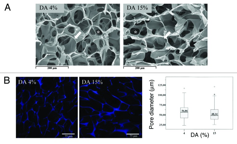
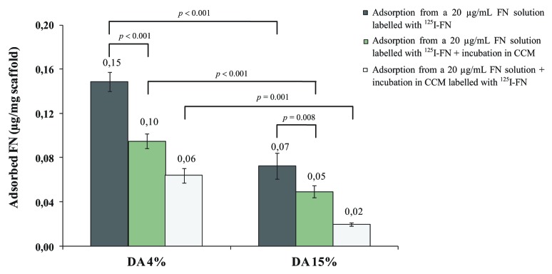
In this study, (125)I-radiolabelling was explored to follow the kinetics and isotherm of fibronectin (FN) adsorption to porous polymeric scaffolds, as well as to assess the elution and exchangeability of pre-adsorbed FN following incubation in serum-containing culture medium. Chitosan (CH) porous scaffolds with two different degrees of acetylation (DA 4% and 15%) were incubated in FN solutions with concentrations ranging from 5 to 50 µg/mL. The kinetic and isotherm of FN adsorption to CH were successfully followed using (125)I-FN as a tracer molecule. While on DA 4% the levels of adsorbed FN increased linearly with FN solution concentration, on DA 15% a saturation plateau was attained, and FN adsorbed amounts were significantly lower. These findings were supported by immunofluorescent studies that revealed, for the same FN solution concentration, higher levels of exposed cell-binding domains on DA 4% as compared with DA 15%. Following incubation in serum containing medium, DA 4% also revealed higher ability to exchange pre-adsorbed FN by new FN molecules from serum than DA 15%. In accordance, when assessing the efficacy of passively adsorbed FN to promote endothelial cell (EC) adhesion to CH, ECs were found to adhere at higher levels to DA 4% as compared with DA 15%, 5 µg/mL of FN being already efficient in promoting cell adhesion and cytoskeletal organization on CH with DA 4%. Taken together the results show that protein radiolabelling can be used as an effective tool to study protein adsorption to porous polymeric scaffolds, both from single and complex protein solutions.

Keywords: 3-D scaffolds; fibronectin; fluorescence; protein adsorption; protein conformation.


Figures

None
Figure 1. Morphological analysis of CH porous scaffolds with DA 4% and 15%. (A) SEM micrographs of transversal cross-sections of the dehydrated scaffolds, showing a highly porous and interconnected structure, with macropores (arrow) and interconnecting pores (*). (B) CLSM imaging of 100 µm thick cryosections of hydrated CH scaffolds. The polymeric structure is shown in blue due to CH autofluorescence upon excitation by the 405 nm laser. The pore diameter distribution resultant from image analysis is shown in the box plot chart, and depicts the following statistics: minimum, first quartile, median, third quartile, and maximum. Pinpoints represent outliers. Values reported correspond to 120 measurements made in CLSM images of cryosections obtained from four different CH scaffolds.
None
Figure 2. Kinetics and isotherms of FN adsorption to CH scaffolds, as determined using 125I-labeled FN. (A) FN adsorption to CH (DA 4%) from a 20 µg/mL FN solution, as a function of incubation time (mean ± SD; n = 8). (B) FN adsorption to CH scaffolds (DA 4 and 15%; 15-h incubation period), as a function of FN solution concentration (mean ± SD; n = 6). All the FN levels shown were determined after subsequent incubation of the porous scaffolds in complete culture medium for 24 h.
None
Figure 3. Fibronectin adsorption to CH scaffolds (DA 4 and 15%) from a 20 µg/mL FN solution, as well as elution and exchangeability of pre-adsorbed FN in the presence of serum proteins. FN adsorption levels were determined by gamma counting after a 15-h incubation period in a 20 µg/mL 125I-labeled FN solution. To estimate the elution of pre-adsorbed FN in the presence of serum proteins, samples were further immersed in complete culture medium (CCM) for 24 h, and the levels of remaining FN quantified by gamma counting. The exchangeability of pre-adsorbed FN by new FN molecules from serum was investigated incubating samples pre-adsorbed with unlabelled FN (20 µg/mL) in CCM containing 125I-FN. Results presented are the mean ± SD (n = 6).
None
Figure 4. Distribution and conformation of FN upon adsorption to CH scaffolds with DA 4% (A) and 15% (B), as probed by immunofluorescent staining of the integrin-binding RGD site of FN. Scaffolds were incubated in a 20 µg/mL FN solution for 15 h, cryosectioned, processed for immunofluorescence, and imaged by CLSM. The scale bar for the low- and high-magnification images corresponds to 300 µm and 75 µm, respectively. (C) Quantitative analysis of exposed cell-binding domains, as determined by fluorimetry. Results reported are the mean ± SD of fluorescence intensity (FI) values correspondent to 8 cryosections.
None
Figure 5. Cell adhesion of HPMEC-ST1.6R cells to CH scaffolds (DA 4 and 15%) previously incubated for 15 h in complete culture medium (CCM) or in FN solutions with concentrations ranging from 5 to 50 µg/mL (mean ± SD; n = 6). TCPS coverslips previously incubated in a 5 µg/mL FN solution were used as 2-D substrate control for EC adhesion. *indicates a significant difference from the corresponding CH samples incubated in FN solutions. δindicates a significant difference from CH samples with DA 4%, when subjected to the same incubation conditions. (p ≤ 0.05).
None
Figure 6. Fluorescent labeling of F-actin (green) and DNA (red) of HPMEC-ST1.6R cells cultured on CH scaffolds (DA 4 and 15%) previously incubated in complete culture medium (A and B) or in FN solutions with concentrations of 5 (C and D) and 50 µg/mL (E and F). Images obtained by CLSM, 144 h after cell seeding. (Scale bar: 200 µm and 50 µm the low- and high-magnification images, respectively).

Similar articles

Cited by

References

    1. Ratner BD, Hoffman AS, Schoen FJ, Lemons JE, eds. Biomaterials science: an introduction to materials in medicine. San Diego: Elsevier Academic Press, 2004.
    1. Chinn JA, Slack SM. Biomaterials: Protein Surface Interactions. In: Bronzino JD, ed. The Biomedical Engineering Handbook. Boca Raton: CRC Press LLC, 2000:1597-608.
    1. Dee KC, Puleo DA, Bizios R. Protein-Surface Interactions. In: Dee KC, Puleo DA, Bizios R, eds. An Introduction To Tissue-Biomaterial Interactions. New York, USA: John Wiley & Sons, Inc, 2003:37-52.
    1. Sousa SR, Moradas-Ferreira P, Saramago B, Melo LV, Barbosa MA. Human serum albumin adsorption on TiO2 from single protein solutions and from plasma. Langmuir. 2004;20:9745–54. doi: 10.1021/la049158d. - DOI - PubMed
    1. González-García C, Sousa SR, Moratal D, Rico P, Salmerón-Sánchez M. Effect of nanoscale topography on fibronectin adsorption, focal adhesion size and matrix organisation. Colloids Surf B Biointerfaces. 2010;77:181–90. doi: 10.1016/j.colsurfb.2010.01.021. - DOI - PubMed

Publication types