

Review
doi: 10.1172/JCI66027.
Epub 2013 May 1.
Hepatic stem cell niches
Affiliations
- PMID: 23635785
- PMCID: PMC3638908
- DOI: 10.1172/JCI66027
Item in Clipboard
Review
Hepatic stem cell niches
J Clin Invest.
2013 May.
Abstract
Stem cell niches are special microenvironments that maintain stem cells and control their behavior to ensure tissue homeostasis and regeneration throughout life. The liver has a high regenerative capacity that involves stem/progenitor cells when the proliferation of hepatocytes is impaired. In recent years progress has been made in the identification of potential hepatic stem cell niches. There is evidence that hepatic progenitor cells can originate from niches in the canals of Hering; in addition, the space of Disse may also serve as a stem cell niche during fetal hematopoiesis and constitute a niche for stellate cells in adults.
Figures
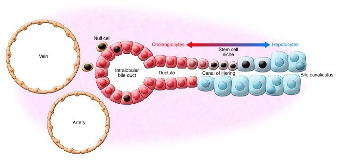
Label-retaining cells (black nuclei) represent putative stem cells and are
located in the canals of Hering as keratin-expressing cells. Also,
cholangiocytes of intralobular bile ducts, small hepatocytes, and
less-characterized null cells retain the BrdU label after acetaminophen
treatment of mice (50). Coexpression
of keratin 19 and albumin in small hepatocytes at the interface of
cholangiocytes and hepatocytes in the canals of Hering indicate that
hepatocytes are generated at this site by stem/progenitor cells (116). A continuous production of
hepatocytes within the portal field is supported by SOX9 fate-mapping
analysis (54). Small cholangiocytes
lining the canals of Hering and ductules may represent cholangiocyte
precursors, which potentially contribute to the cholangiocyte population of
larger bile ducts (116) and could
explain the presence of label-retaining cholangiocytes at this site (50).
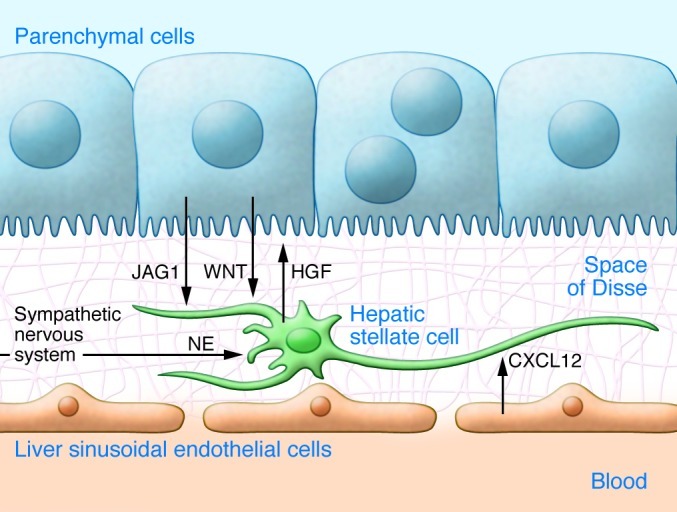
Hepatic stellate cells (HSCs) are located on basement membrane proteins such
as laminin and collagen type IV (grid) between LSECs and parenchymal cells
(PCs). HSCs are attracted to LSECs through CXCL12 and are associated with
PCs, which express Notch ligands such as jagged 1 (JAG1) and release
canonical WNT ligands to influence stellate cell development (72). Quiescent stellate cells in turn
secrete HGF, which is probably involved in liver tissue homeostasis and can
support hepatic progenitor cells (75,
76). HSCs receive signals from
the sympathetic nervous system through norepinephrine (NE) release (112, 115).
Similar articles
-
The diversity and plasticity of adult hepatic progenitor cells and their niche.Liver Int. 2017 Sep;37(9):1260-1271. doi: 10.1111/liv.13377. Epub 2017 Feb 23. Liver Int. 2017. PMID: 28135758 Free PMC article. Review.
-
Space of Disse: a stem cell niche in the liver.Biol Chem. 2019 Dec 18;401(1):81-95. doi: 10.1515/hsz-2019-0283. Biol Chem. 2019. PMID: 31318687 Review.
-
Contribution of Resident Stem Cells to Liver and Biliary Tree Regeneration in Human Diseases.Int J Mol Sci. 2018 Sep 25;19(10):2917. doi: 10.3390/ijms19102917. Int J Mol Sci. 2018. PMID: 30257529 Free PMC article. Review.
-
Hepatic stem cells and liver development.Methods Mol Biol. 2010;640:181-236. doi: 10.1007/978-1-60761-688-7_10. Methods Mol Biol. 2010. PMID: 20645053
-
Founder cells for hepatocytes during liver regeneration: from identification to application.Cell Mol Life Sci. 2020 Aug;77(15):2887-2898. doi: 10.1007/s00018-020-03457-3. Epub 2020 Feb 14. Cell Mol Life Sci. 2020. PMID: 32060582 Free PMC article. Review.
Cited by
-
The Role of Embryonic Stem Cell-expressed RAS (ERAS) in the Maintenance of Quiescent Hepatic Stellate Cells.J Biol Chem. 2016 Apr 15;291(16):8399-413. doi: 10.1074/jbc.M115.700088. Epub 2016 Feb 16. J Biol Chem. 2016. PMID: 26884329 Free PMC article.
-
Bile acid signaling and liver regeneration.Biochim Biophys Acta. 2015 Feb;1849(2):196-200. doi: 10.1016/j.bbagrm.2014.05.021. Epub 2014 May 27. Biochim Biophys Acta. 2015. PMID: 24878541 Free PMC article. Review.
-
Macrophages and Stem Cells-Two to Tango for Tissue Repair?Biomolecules. 2021 May 6;11(5):697. doi: 10.3390/biom11050697. Biomolecules. 2021. PMID: 34066618 Free PMC article. Review.
-
A Protocol for the Isolation of Oval Cells without Preconditioning.Int J Mol Sci. 2024 Sep 29;25(19):10497. doi: 10.3390/ijms251910497. Int J Mol Sci. 2024. PMID: 39408831 Free PMC article.
-
Cytochrome P450 1B1: An unexpected modulator of liver fatty acid homeostasis.Arch Biochem Biophys. 2015 Apr 1;571:21-39. doi: 10.1016/j.abb.2015.02.010. Epub 2015 Feb 20. Arch Biochem Biophys. 2015. PMID: 25703193 Free PMC article.
References
-
- Schofield R. The relationship between the spleen colony-forming cell and the haemopoietic stem cell. Blood Cells. 1978;4(1–2):7–25. - PubMed
Publication types
MeSH terms
LinkOut - more resources
Full Text Sources
Other Literature Sources
Medical
