Review
. 2013 May;123(5):1887-901.
doi: 10.1172/JCI66028. Epub 2013 May 1.

Evolving therapies for liver fibrosis

Affiliations
Review

Evolving therapies for liver fibrosis

Detlef Schuppan et al. J Clin Invest. 2013 May.

Abstract

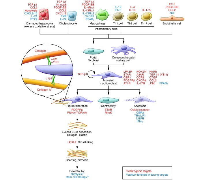
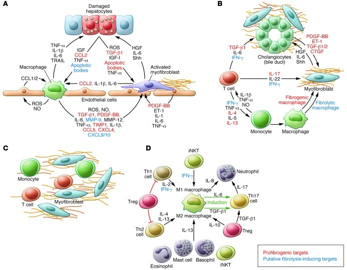
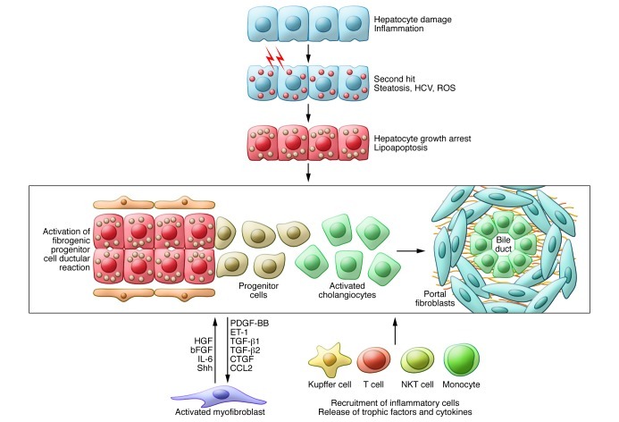
Fibrosis is an intrinsic response to chronic injury, maintaining organ integrity when extensive necrosis or apoptosis occurs. With protracted damage, fibrosis can progress toward excessive scarring and organ failure, as in liver cirrhosis. To date, antifibrotic treatment of fibrosis represents an unconquered area for drug development, with enormous potential but also high risks. Preclinical research has yielded numerous targets for antifibrotic agents, some of which have entered early-phase clinical studies, but progress has been hampered due to the relative lack of sensitive and specific biomarkers to measure fibrosis progression or reversal. Here we focus on antifibrotic approaches for liver that address specific cell types and functional units that orchestrate fibrotic wound healing responses and have a sound preclinical database or antifibrotic activity in early clinical trials. We also touch upon relevant clinical study endpoints, optimal study design, and developments in fibrosis imaging and biomarkers.


Figures

Figure 1
Figure 1. Myofibroblasts and their fibrogenic activation.
Cells and major factors upstream of quiescent portal fibroblasts and hepatic stellate cells that induce transformation to fibrogenic myofibroblasts. This schematic highlights several major targets to treat liver fibrosis. Notably, the ECM itself can serve as modulator of fibrogenesis and fibrolysis. Thus collagen fibrils become crosslinked by LOXL2, which contributes to the reduced reversibility of advanced fibrosis, and collagen-binding ECM receptors (especially the integrins α1β1, α2β1, and α11β1) confer signals of stress or stress relaxation that either maintain fibrogenic activation or induce fibrolytic activity of the myofibroblasts. Additional minor contributors to fibrogenic activation are not shown here (see text for details). A2AR, adenosine 2A receptor; AT1R, angiotensin 1 receptor; CBR1, cannabinoid receptor 1; ET-1, endothelin-1; ETAR, endothelin A receptor; FXR, farnesoid X receptor; Hh(R), hedgehog (receptor); Int, integrin; LPA1R, lysophosphatidic acid receptor 1; NGFR, nerve growth factor receptor; PTX2, pentraxin 2; TRAILR, TNF-related apoptosis-inducing ligand receptor; YB-1, Y-box binding protein.
Figure 2
Figure 2. Multicellular context of fibrogenesis and fibrolysis.
Shown are the postulated major cellular functional units and secreted factors that should be addressed in their complexity when designing effective antifibrotic strategies. (A) Vascular unit. (B) Biliary unit. (C) Inflammatory unit. (D) Cells and factors that affect macrophage polarization. Macrophages (and monocytes as macrophage precursors) are major modulators of inflammation and tissue remodeling. Cells and factors that induce either M1 or M2 polarization are also linked to the generation of fibrogenic Th17 cells and neutrophil recruitment. See text for details. B and C highlight factors not shown in A and B, respectively. Baso, basophil; EO, eosinophil; Mast, mast cell; PMN, polymorphonuclear neutrophil; TIMP, tissue inhibitor of metalloproteinases.
Figure 3
Figure 3. Activated cholangiocytes as drivers of fibrosis progression.
Activated cholangiocytes are related, if not identical, to biliary progenitor cells. These cells proliferate in active biliary diseases and during massive hepatocyte growth arrest or apoptosis, as in severe NASH, ASH, or viral hepatitis. Biliary progenitor cells are regularly found in more advanced fibrosis (especially Metavir stage F2 or higher). They replicate ductal plate formation by induction of a portal fibrotic matrix via secretion of profibrogenic factors and recruitment and activation of myofibroblasts, and also Kupffer cells and monocytes and other inflammatory cells like T and NKT cells. The recruited myofibroblasts (and the inflammatory cells) secrete factors and ECM components that maintain these fibrogenic units and support their differentiation into more mature biliary structures that are embedded in a collagen-rich ECM.

References

    1. Friedman SL. Mechanisms of hepatic fibrogenesis. Gastroenterology. 2008;134(6):1655–1669. doi: 10.1053/j.gastro.2008.03.003. - DOI - PMC - PubMed
    1. Schuppan D, Afdhal NH. Liver cirrhosis. Lancet. 2008;371(9615):838–851. doi: 10.1016/S0140-6736(08)60383-9. - DOI - PMC - PubMed
    1. Gao B, Bataller R. Alcoholic liver disease: pathogenesis and new therapeutic targets. Gastroenterology. 2011;141(5):1572–1585. doi: 10.1053/j.gastro.2011.09.002. - DOI - PMC - PubMed
    1. Tilg H, Moschen AR. Evolution of inflammation in nonalcoholic fatty liver disease: the multiple parallel hits hypothesis. Hepatology. 2010;52(5):1836–1846. doi: 10.1002/hep.24001. - DOI - PubMed
    1. Bohinc BN, Diehl AM. Mechanisms of disease progression in NASH: new paradigms. Clin Liver Dis. 2012;16(3):549–565. doi: 10.1016/j.cld.2012.05.002. - DOI - PubMed

Publication types